An official website of the United States government
The .gov means it's official. Federal government websites often end in .gov or .mil. Before sharing sensitive information, make sure you're on a federal government site.
The site is secure. The https:// ensures that you are connecting to the official website and that any information you provide is encrypted and transmitted securely.
- Publications
- Account settings
- Browse Titles
NCBI Bookshelf. A service of the National Library of Medicine, National Institutes of Health.
StatPearls [Internet]. Treasure Island (FL): StatPearls Publishing; 2024 Jan-.


StatPearls [Internet].
Case control studies.
Steven Tenny ; Connor C. Kerndt ; Mary R. Hoffman .
Affiliations
Last Update: March 27, 2023 .
- Introduction
A case-control study is a type of observational study commonly used to look at factors associated with diseases or outcomes. [1] The case-control study starts with a group of cases, which are the individuals who have the outcome of interest. The researcher then tries to construct a second group of individuals called the controls, who are similar to the case individuals but do not have the outcome of interest. The researcher then looks at historical factors to identify if some exposure(s) is/are found more commonly in the cases than the controls. If the exposure is found more commonly in the cases than in the controls, the researcher can hypothesize that the exposure may be linked to the outcome of interest.
For example, a researcher may want to look at the rare cancer Kaposi's sarcoma. The researcher would find a group of individuals with Kaposi's sarcoma (the cases) and compare them to a group of patients who are similar to the cases in most ways but do not have Kaposi's sarcoma (controls). The researcher could then ask about various exposures to see if any exposure is more common in those with Kaposi's sarcoma (the cases) than those without Kaposi's sarcoma (the controls). The researcher might find that those with Kaposi's sarcoma are more likely to have HIV, and thus conclude that HIV may be a risk factor for the development of Kaposi's sarcoma.
There are many advantages to case-control studies. First, the case-control approach allows for the study of rare diseases. If a disease occurs very infrequently, one would have to follow a large group of people for a long period of time to accrue enough incident cases to study. Such use of resources may be impractical, so a case-control study can be useful for identifying current cases and evaluating historical associated factors. For example, if a disease developed in 1 in 1000 people per year (0.001/year) then in ten years one would expect about 10 cases of a disease to exist in a group of 1000 people. If the disease is much rarer, say 1 in 1,000,0000 per year (0.0000001/year) this would require either having to follow 1,000,0000 people for ten years or 1000 people for 1000 years to accrue ten total cases. As it may be impractical to follow 1,000,000 for ten years or to wait 1000 years for recruitment, a case-control study allows for a more feasible approach.
Second, the case-control study design makes it possible to look at multiple risk factors at once. In the example above about Kaposi's sarcoma, the researcher could ask both the cases and controls about exposures to HIV, asbestos, smoking, lead, sunburns, aniline dye, alcohol, herpes, human papillomavirus, or any number of possible exposures to identify those most likely associated with Kaposi's sarcoma.
Case-control studies can also be very helpful when disease outbreaks occur, and potential links and exposures need to be identified. This study mechanism can be commonly seen in food-related disease outbreaks associated with contaminated products, or when rare diseases start to increase in frequency, as has been seen with measles in recent years.
Because of these advantages, case-control studies are commonly used as one of the first studies to build evidence of an association between exposure and an event or disease.
In a case-control study, the investigator can include unequal numbers of cases with controls such as 2:1 or 4:1 to increase the power of the study.
Disadvantages and Limitations
The most commonly cited disadvantage in case-control studies is the potential for recall bias. [2] Recall bias in a case-control study is the increased likelihood that those with the outcome will recall and report exposures compared to those without the outcome. In other words, even if both groups had exactly the same exposures, the participants in the cases group may report the exposure more often than the controls do. Recall bias may lead to concluding that there are associations between exposure and disease that do not, in fact, exist. It is due to subjects' imperfect memories of past exposures. If people with Kaposi's sarcoma are asked about exposure and history (e.g., HIV, asbestos, smoking, lead, sunburn, aniline dye, alcohol, herpes, human papillomavirus), the individuals with the disease are more likely to think harder about these exposures and recall having some of the exposures that the healthy controls.
Case-control studies, due to their typically retrospective nature, can be used to establish a correlation between exposures and outcomes, but cannot establish causation . These studies simply attempt to find correlations between past events and the current state.
When designing a case-control study, the researcher must find an appropriate control group. Ideally, the case group (those with the outcome) and the control group (those without the outcome) will have almost the same characteristics, such as age, gender, overall health status, and other factors. The two groups should have similar histories and live in similar environments. If, for example, our cases of Kaposi's sarcoma came from across the country but our controls were only chosen from a small community in northern latitudes where people rarely go outside or get sunburns, asking about sunburn may not be a valid exposure to investigate. Similarly, if all of the cases of Kaposi's sarcoma were found to come from a small community outside a battery factory with high levels of lead in the environment, then controls from across the country with minimal lead exposure would not provide an appropriate control group. The investigator must put a great deal of effort into creating a proper control group to bolster the strength of the case-control study as well as enhance their ability to find true and valid potential correlations between exposures and disease states.
Similarly, the researcher must recognize the potential for failing to identify confounding variables or exposures, introducing the possibility of confounding bias, which occurs when a variable that is not being accounted for that has a relationship with both the exposure and outcome. This can cause us to accidentally be studying something we are not accounting for but that may be systematically different between the groups.
The major method for analyzing results in case-control studies is the odds ratio (OR). The odds ratio is the odds of having a disease (or outcome) with the exposure versus the odds of having the disease without the exposure. The most straightforward way to calculate the odds ratio is with a 2 by 2 table divided by exposure and disease status (see below). Mathematically we can write the odds ratio as follows.
Odds ratio = [(Number exposed with disease)/(Number exposed without disease) ]/[(Number not exposed to disease)/(Number not exposed without disease) ]
This can be rewritten as:
Odds ratio = [ (Number exposed with disease) x (Number not exposed without disease) ] / [ (Number exposed without disease ) x (Number not exposed with disease) ]
The odds ratio tells us how strongly the exposure is related to the disease state. An odds ratio of greater than one implies the disease is more likely with exposure. An odds ratio of less than one implies the disease is less likely with exposure and thus the exposure may be protective. For example, a patient with a prior heart attack taking a daily aspirin has a decreased odds of having another heart attack (odds ratio less than one). An odds ratio of one implies there is no relation between the exposure and the disease process.
Odds ratios are often confused with Relative Risk (RR), which is a measure of the probability of the disease or outcome in the exposed vs unexposed groups. For very rare conditions, the OR and RR may be very similar, but they are measuring different aspects of the association between outcome and exposure. The OR is used in case-control studies because RR cannot be estimated; whereas in randomized clinical trials, a direct measurement of the development of events in the exposed and unexposed groups can be seen. RR is also used to compare risk in other prospective study designs.
- Issues of Concern
The main issues of concern with a case-control study are recall bias, its retrospective nature, the need for a careful collection of measured variables, and the selection of an appropriate control group. [3] These are discussed above in the disadvantages section.
- Clinical Significance
A case-control study is a good tool for exploring risk factors for rare diseases or when other study types are not feasible. Many times an investigator will hypothesize a list of possible risk factors for a disease process and will then use a case-control study to see if there are any possible associations between the risk factors and the disease process. The investigator can then use the data from the case-control study to focus on a few of the most likely causative factors and develop additional hypotheses or questions. Then through further exploration, often using other study types (such as cohort studies or randomized clinical studies) the researcher may be able to develop further support for the evidence of the possible association between the exposure and the outcome.
- Enhancing Healthcare Team Outcomes
Case-control studies are prevalent in all fields of medicine from nursing and pharmacy to use in public health and surgical patients. Case-control studies are important for each member of the health care team to not only understand their common occurrence in research but because each part of the health care team has parts to contribute to such studies. One of the most important things each party provides is helping identify correct controls for the cases. Matching the controls across a spectrum of factors outside of the elements of interest take input from nurses, pharmacists, social workers, physicians, demographers, and more. Failure for adequate selection of controls can lead to invalid study conclusions and invalidate the entire study.
- Review Questions
- Access free multiple choice questions on this topic.
- Comment on this article.
2x2 table with calculations for the odds ratio and 95% confidence interval for the odds ratio Contributed by Steven Tenny MD, MPH, MBA
Disclosure: Steven Tenny declares no relevant financial relationships with ineligible companies.
Disclosure: Connor Kerndt declares no relevant financial relationships with ineligible companies.
Disclosure: Mary Hoffman declares no relevant financial relationships with ineligible companies.
This book is distributed under the terms of the Creative Commons Attribution-NonCommercial-NoDerivatives 4.0 International (CC BY-NC-ND 4.0) ( http://creativecommons.org/licenses/by-nc-nd/4.0/ ), which permits others to distribute the work, provided that the article is not altered or used commercially. You are not required to obtain permission to distribute this article, provided that you credit the author and journal.
- Cite this Page Tenny S, Kerndt CC, Hoffman MR. Case Control Studies. [Updated 2023 Mar 27]. In: StatPearls [Internet]. Treasure Island (FL): StatPearls Publishing; 2024 Jan-.
In this Page
Bulk download.
- Bulk download StatPearls data from FTP
Related information
- PMC PubMed Central citations
- PubMed Links to PubMed
Similar articles in PubMed
- Suicidal Ideation. [StatPearls. 2024] Suicidal Ideation. Harmer B, Lee S, Rizvi A, Saadabadi A. StatPearls. 2024 Jan
- Qualitative Study. [StatPearls. 2024] Qualitative Study. Tenny S, Brannan JM, Brannan GD. StatPearls. 2024 Jan
- Folic acid supplementation and malaria susceptibility and severity among people taking antifolate antimalarial drugs in endemic areas. [Cochrane Database Syst Rev. 2022] Folic acid supplementation and malaria susceptibility and severity among people taking antifolate antimalarial drugs in endemic areas. Crider K, Williams J, Qi YP, Gutman J, Yeung L, Mai C, Finkelstain J, Mehta S, Pons-Duran C, Menéndez C, et al. Cochrane Database Syst Rev. 2022 Feb 1; 2(2022). Epub 2022 Feb 1.
- Review The epidemiology of classic, African, and immunosuppressed Kaposi's sarcoma. [Epidemiol Rev. 1991] Review The epidemiology of classic, African, and immunosuppressed Kaposi's sarcoma. Wahman A, Melnick SL, Rhame FS, Potter JD. Epidemiol Rev. 1991; 13:178-99.
- Review Epidemiology of Kaposi's sarcoma. [Cancer Surv. 1991] Review Epidemiology of Kaposi's sarcoma. Beral V. Cancer Surv. 1991; 10:5-22.
Recent Activity
- Case Control Studies - StatPearls Case Control Studies - StatPearls
Your browsing activity is empty.
Activity recording is turned off.
Turn recording back on
Connect with NLM
National Library of Medicine 8600 Rockville Pike Bethesda, MD 20894
Web Policies FOIA HHS Vulnerability Disclosure
Help Accessibility Careers
Study Design 101: Case Control Study
- Case Report
- Case Control Study
- Cohort Study
- Randomized Controlled Trial
- Practice Guideline
- Systematic Review
- Meta-Analysis
- Helpful Formulas
- Finding Specific Study Types
A study that compares patients who have a disease or outcome of interest (cases) with patients who do not have the disease or outcome (controls), and looks back retrospectively to compare how frequently the exposure to a risk factor is present in each group to determine the relationship between the risk factor and the disease.
Case control studies are observational because no intervention is attempted and no attempt is made to alter the course of the disease. The goal is to retrospectively determine the exposure to the risk factor of interest from each of the two groups of individuals: cases and controls. These studies are designed to estimate odds.
Case control studies are also known as "retrospective studies" and "case-referent studies."
- Good for studying rare conditions or diseases
- Less time needed to conduct the study because the condition or disease has already occurred
- Lets you simultaneously look at multiple risk factors
- Useful as initial studies to establish an association
- Can answer questions that could not be answered through other study designs
Disadvantages
- Retrospective studies have more problems with data quality because they rely on memory and people with a condition will be more motivated to recall risk factors (also called recall bias).
- Not good for evaluating diagnostic tests because it's already clear that the cases have the condition and the controls do not
- It can be difficult to find a suitable control group
Design pitfalls to look out for
Care should be taken to avoid confounding, which arises when an exposure and an outcome are both strongly associated with a third variable. Controls should be subjects who might have been cases in the study but are selected independent of the exposure. Cases and controls should also not be "over-matched."
Is the control group appropriate for the population? Does the study use matching or pairing appropriately to avoid the effects of a confounding variable? Does it use appropriate inclusion and exclusion criteria?
Fictitious Example
There is a suspicion that zinc oxide, the white non-absorbent sunscreen traditionally worn by lifeguards is more effective at preventing sunburns that lead to skin cancer than absorbent sunscreen lotions. A case-control study was conducted to investigate if exposure to zinc oxide is a more effective skin cancer prevention measure. The study involved comparing a group of former lifeguards that had developed cancer on their cheeks and noses (cases) to a group of lifeguards without this type of cancer (controls) and assess their prior exposure to zinc oxide or absorbent sunscreen lotions.
This study would be retrospective in that the former lifeguards would be asked to recall which type of sunscreen they used on their face and approximately how often. This could be either a matched or unmatched study, but efforts would need to be made to ensure that the former lifeguards are of the same average age, and lifeguarded for a similar number of seasons and amount of time per season.
Real-life Examples
Boubekri, M., Cheung, I., Reid, K., Wang, C., & Zee, P. (2014). Impact of windows and daylight exposure on overall health and sleep quality of office workers: a case-control pilot study. Journal of Clinical Sleep Medicine : JCSM : Official Publication of the American Academy of Sleep Medicine, 10 (6), 603-611. https://doi.org/10.5664/jcsm.3780
This pilot study explored the impact of exposure to daylight on the health of office workers (measuring well-being and sleep quality subjectively, and light exposure, activity level and sleep-wake patterns via actigraphy). Individuals with windows in their workplaces had more light exposure, longer sleep duration, and more physical activity. They also reported a better scores in the areas of vitality and role limitations due to physical problems, better sleep quality and less sleep disturbances.
Togha, M., Razeghi Jahromi, S., Ghorbani, Z., Martami, F., & Seifishahpar, M. (2018). Serum Vitamin D Status in a Group of Migraine Patients Compared With Healthy Controls: A Case-Control Study. Headache, 58 (10), 1530-1540. https://doi.org/10.1111/head.13423
This case-control study compared serum vitamin D levels in individuals who experience migraine headaches with their matched controls. Studied over a period of thirty days, individuals with higher levels of serum Vitamin D was associated with lower odds of migraine headache.
Related Formulas
- Odds ratio in an unmatched study
- Odds ratio in a matched study
Related Terms
A patient with the disease or outcome of interest.
Confounding
When an exposure and an outcome are both strongly associated with a third variable.
A patient who does not have the disease or outcome.
Matched Design
Each case is matched individually with a control according to certain characteristics such as age and gender. It is important to remember that the concordant pairs (pairs in which the case and control are either both exposed or both not exposed) tell us nothing about the risk of exposure separately for cases or controls.
Observed Assignment
The method of assignment of individuals to study and control groups in observational studies when the investigator does not intervene to perform the assignment.
Unmatched Design
The controls are a sample from a suitable non-affected population.
Now test yourself!
1. Case Control Studies are prospective in that they follow the cases and controls over time and observe what occurs.
a) True b) False
2. Which of the following is an advantage of Case Control Studies?
a) They can simultaneously look at multiple risk factors. b) They are useful to initially establish an association between a risk factor and a disease or outcome. c) They take less time to complete because the condition or disease has already occurred. d) b and c only e) a, b, and c
Evidence Pyramid - Navigation
- Meta- Analysis
- Case Reports
- << Previous: Case Report
- Next: Cohort Study >>

- Last Updated: Sep 25, 2023 10:59 AM
- URL: https://guides.himmelfarb.gwu.edu/studydesign101

- Himmelfarb Intranet
- Privacy Notice
- Terms of Use
- GW is committed to digital accessibility. If you experience a barrier that affects your ability to access content on this page, let us know via the Accessibility Feedback Form .
- Himmelfarb Health Sciences Library
- 2300 Eye St., NW, Washington, DC 20037
- Phone: (202) 994-2850
- [email protected]
- https://himmelfarb.gwu.edu
- - Google Chrome
Intended for healthcare professionals
- Access provided by Google Indexer
- My email alerts
- BMA member login
- Username * Password * Forgot your log in details? Need to activate BMA Member Log In Log in via OpenAthens Log in via your institution

Search form
- Advanced search
- Search responses
- Search blogs
- Nested case-control...
Nested case-control studies: advantages and disadvantages
- Related content
- Peer review
- Philip Sedgwick , reader in medical statistics and medical education 1
- 1 Centre for Medical and Healthcare Education, St George’s, University of London, London, UK
- p.sedgwick{at}sgul.ac.uk
Researchers investigated whether antipsychotic drugs were associated with venous thromboembolism. A population based nested case-control study design was used. Data were taken from the UK QResearch primary care database consisting of 7 267 673 patients. Cases were adult patients with a first ever record of venous thromboembolism between 1 January 1996 and 1 July 2007. For each case, up to four controls were identified, matched by age, calendar time, sex, and practice. Exposure to antipsychotic drugs was assessed on the basis of prescriptions on, or during the 24 months before, the index date. 1
There were 25 532 eligible cases (15 975 with deep vein thrombosis and 9557 with pulmonary embolism) and 89 491 matched controls. The primary outcome was the odds ratios for venous thromboembolism associated with antipsychotic drugs adjusted for comorbidity and concomitant drug exposure. When adjusted using logistic regression to control for potential confounding, prescription of antipsychotic drugs in the previous 24 months was significantly associated with an increased occurrence of venous thromboembolism compared with non-use (odds ratio 1.32, 95% confidence interval 1.23 to 1.42). The researchers concluded that prescription of antipsychotic drugs was associated with venous thromboembolism in a large primary care population.
Which of the following statements, if any, are true?
a) The nested case-control study is a retrospective design
b) The study design minimised selection bias compared with a case-control study
c) Recall bias was minimised compared with a case-control study
d) Causality could be inferred from the association between prescription of antipsychotic drugs and venous thromboembolism
Statements a , b , and c are true, whereas d is false.
The aim of the study was to investigate whether prescription of antipsychotic drugs was associated with venous thromboembolism. A nested case-control study design was used. The study design was an observational one that incorporated the concept of the traditional case-control study within an established cohort. This design overcomes some of the disadvantages associated with case-control studies, 2 while incorporating some of the advantages of cohort studies. 3 4
Data for the study above were extracted from the UK QResearch primary care database, a computerised register of anonymised longitudinal medical records for patients registered at more than 500 UK general practices. Patient data were recorded prospectively, the database having been updated regularly as patients visited their GP. Cases were all adult patients in the register with a first ever record of venous thromboembolism between 1 January 1996 and 1 July 2007. There were 25 532 cases in total. For each case, up to four controls were identified from the register, matched by age, calendar time, sex, and practice. In total, 89 491 matched controls were obtained. Data relating to prescriptions for antipsychotic drugs on, or during the 24 months before, the index date were extracted for the cases and controls. The index date was the date in the register when venous thromboembolism was recorded for the case. The cases and controls were compared to ascertain whether exposure to prescription of antipsychotic drugs was more common in one group than in the other. Despite the data for the cases and controls being collected prospectively, the nested case-control study is described as retrospective ( a is true) because it involved looking back at events that had already taken place and been recorded in the register.
Selection bias is of particular concern in the traditional case-control study. Described in a previous question, 5 selection bias is the systematic difference between the study participants and the population they are meant to represent with respect to their characteristics, including demographics and morbidity. Cases and controls are often selected through convenience sampling. Cases are typically recruited from hospitals or general practices because they are convenient and easily accessible to researchers. Controls are often recruited from the same hospital clinics or general practices as the cases. Therefore, the selected cases may not be representative of the population of all cases. Equally, the controls might not be representative of otherwise healthy members of the population. The above nested case-control study was population based, with the QResearch primary care database incorporating a large proportion of the UK population. The cases and controls were selected from the database and therefore should be more representative of the population than those in a traditional case-control study. Hence, selection bias was minimised by using the nested case-control study design ( b is true).
The traditional case-control study involves participants recalling information about past exposure to risk factors after identification as a case or control. The study design is prone to recall bias, as described in a previous question. 6 Recall bias is the systematic difference between cases and controls in the accuracy of information recalled. Recall bias will exist if participants have selective preconceptions about the association between the disease and past exposure to the risk factor(s). Cases may, for example, recall information more accurately than controls, possibly because of an association with the disease or outcome. Although in the study above the cases and controls were identified retrospectively, the data for the QResearch primary care database were collected prospectively. Therefore, there was no reason for any systematic differences between groups of study participants in the accuracy of the information collected. Therefore, recall bias was minimised compared with a traditional case-control study ( c is true).
Not all of the patient records in the UK QResearch primary care database were used to explore the association between prescription of antipsychotic drugs and development of venous thromboembolism. A nested case-control study was used instead, with cases and controls matched on age, calendar time, sex, and practice. This was because it was statistically more efficient to control for the effects of age, calendar time, sex, and practice by matching cases and controls on these variables at the design stage, rather than controlling for their potential confounding effects when the data were analysed. The matching variables were considered to be important factors that could potentially confound the association between prescription of antipsychotic drugs and venous thromboembolism, but they were not of interest as potential risk factors in themselves. Matching in case-control studies has been described in a previous question. 7
Unlike a traditional case-control study, the data in the example above were recorded prospectively. Therefore, it was possible to determine whether prescription of antipsychotic drugs preceded the occurrence of venous thromboembolism. Nonetheless, only association, and not causation, can be inferred from the results of the above nested case-control study ( d is false)—that is, those people who were exposed to prescribed antipsychotic drugs were more likely to have developed venous thromboembolism. This is because the observed association between prescribed antipsychotic drugs and occurrence of venous thromboembolism may have been due to confounding. In particular, it was not possible to measure and then control for, through statistical analysis, all factors that may have affected the occurrence of venous thromboembolism.
The example above is typical of a nested case-control study; the health records for a group of patients that have already been collected and stored in an electronic database are used to explore the association between one or more risk factors and a disease or condition. The management of such databases means it is possible for a variety of studies to be undertaken, each investigating the risk factors associated with different diseases or outcomes. Nested case-control studies are therefore relatively inexpensive to perform. However, the major disadvantage of nested case-control studies is that not all pertinent risk factors are likely to have been recorded. Furthermore, because many different healthcare professionals will be involved in patient care, risk factors and outcome(s) will probably not have been measured with the same accuracy and consistency throughout. It may also be problematic if the diagnosis of the disease or outcome changes with time.
Cite this as: BMJ 2014;348:g1532
Competing interests: None declared.
- ↵ Parker C, Coupland C, Hippisley-Cox J. Antipsychotic drugs and risk of venous thromboembolism: nested case-control study. BMJ 2010 ; 341 : c4245 . OpenUrl Abstract / FREE Full Text
- ↵ Sedgwick P. Case-control studies: advantages and disadvantages. BMJ 2014 ; 348 : f7707 . OpenUrl CrossRef
- ↵ Sedgwick P. Prospective cohort studies: advantages and disadvantages. BMJ 2013 ; 347 : f6726 . OpenUrl FREE Full Text
- ↵ Sedgwick P. Retrospective cohort studies: advantages and disadvantages. BMJ 2014 ; 348 : g1072 . OpenUrl FREE Full Text
- ↵ Sedgwick P. Selection bias versus allocation bias. BMJ 2013 ; 346 : f3345 . OpenUrl FREE Full Text
- ↵ Sedgwick P. What is recall bias? BMJ 2012 ; 344 : e3519 . OpenUrl FREE Full Text
- ↵ Sedgwick P. Why match in case-control studies? BMJ 2012 ; 344 : e691 . OpenUrl FREE Full Text
What Is A Case Control Study?
Julia Simkus
Editor at Simply Psychology
BA (Hons) Psychology, Princeton University
Julia Simkus is a graduate of Princeton University with a Bachelor of Arts in Psychology. She is currently studying for a Master's Degree in Counseling for Mental Health and Wellness in September 2023. Julia's research has been published in peer reviewed journals.
Learn about our Editorial Process
Saul Mcleod, PhD
Editor-in-Chief for Simply Psychology
BSc (Hons) Psychology, MRes, PhD, University of Manchester
Saul Mcleod, PhD., is a qualified psychology teacher with over 18 years of experience in further and higher education. He has been published in peer-reviewed journals, including the Journal of Clinical Psychology.
Olivia Guy-Evans, MSc
Associate Editor for Simply Psychology
BSc (Hons) Psychology, MSc Psychology of Education
Olivia Guy-Evans is a writer and associate editor for Simply Psychology. She has previously worked in healthcare and educational sectors.
On This Page:
A case-control study is a research method where two groups of people are compared – those with the condition (cases) and those without (controls). By looking at their past, researchers try to identify what factors might have contributed to the condition in the ‘case’ group.
Explanation
A case-control study looks at people who already have a certain condition (cases) and people who don’t (controls). By comparing these two groups, researchers try to figure out what might have caused the condition. They look into the past to find clues, like habits or experiences, that are different between the two groups.
The “cases” are the individuals with the disease or condition under study, and the “controls” are similar individuals without the disease or condition of interest.
The controls should have similar characteristics (i.e., age, sex, demographic, health status) to the cases to mitigate the effects of confounding variables .
Case-control studies identify any associations between an exposure and an outcome and help researchers form hypotheses about a particular population.
Researchers will first identify the two groups, and then look back in time to investigate which subjects in each group were exposed to the condition.
If the exposure is found more commonly in the cases than the controls, the researcher can hypothesize that the exposure may be linked to the outcome of interest.
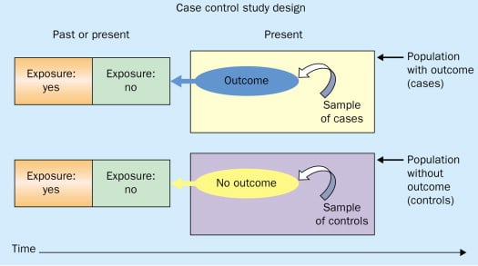
Figure: Schematic diagram of case-control study design. Kenneth F. Schulz and David A. Grimes (2002) Case-control studies: research in reverse . The Lancet Volume 359, Issue 9304, 431 – 434
Quick, inexpensive, and simple
Because these studies use already existing data and do not require any follow-up with subjects, they tend to be quicker and cheaper than other types of research. Case-control studies also do not require large sample sizes.
Beneficial for studying rare diseases
Researchers in case-control studies start with a population of people known to have the target disease instead of following a population and waiting to see who develops it. This enables researchers to identify current cases and enroll a sufficient number of patients with a particular rare disease.
Useful for preliminary research
Case-control studies are beneficial for an initial investigation of a suspected risk factor for a condition. The information obtained from cross-sectional studies then enables researchers to conduct further data analyses to explore any relationships in more depth.
Limitations
Subject to recall bias.
Participants might be unable to remember when they were exposed or omit other details that are important for the study. In addition, those with the outcome are more likely to recall and report exposures more clearly than those without the outcome.
Difficulty finding a suitable control group
It is important that the case group and the control group have almost the same characteristics, such as age, gender, demographics, and health status.
Forming an accurate control group can be challenging, so sometimes researchers enroll multiple control groups to bolster the strength of the case-control study.
Do not demonstrate causation
Case-control studies may prove an association between exposures and outcomes, but they can not demonstrate causation.
A case-control study is an observational study where researchers analyzed two groups of people (cases and controls) to look at factors associated with particular diseases or outcomes.
Below are some examples of case-control studies:
- Investigating the impact of exposure to daylight on the health of office workers (Boubekri et al., 2014).
- Comparing serum vitamin D levels in individuals who experience migraine headaches with their matched controls (Togha et al., 2018).
- Analyzing correlations between parental smoking and childhood asthma (Strachan and Cook, 1998).
- Studying the relationship between elevated concentrations of homocysteine and an increased risk of vascular diseases (Ford et al., 2002).
- Assessing the magnitude of the association between Helicobacter pylori and the incidence of gastric cancer (Helicobacter and Cancer Collaborative Group, 2001).
- Evaluating the association between breast cancer risk and saturated fat intake in postmenopausal women (Howe et al., 1990).
Frequently asked questions
1. what’s the difference between a case-control study and a cross-sectional study.
Case-control studies are different from cross-sectional studies in that case-control studies compare groups retrospectively while cross-sectional studies analyze information about a population at a specific point in time.
In cross-sectional studies , researchers are simply examining a group of participants and depicting what already exists in the population.
2. What’s the difference between a case-control study and a longitudinal study?
Case-control studies compare groups retrospectively, while longitudinal studies can compare groups either retrospectively or prospectively.
In a longitudinal study , researchers monitor a population over an extended period of time, and they can be used to study developmental shifts and understand how certain things change as we age.
In addition, case-control studies look at a single subject or a single case, whereas longitudinal studies can be conducted on a large group of subjects.
3. What’s the difference between a case-control study and a retrospective cohort study?
Case-control studies are retrospective as researchers begin with an outcome and trace backward to investigate exposure; however, they differ from retrospective cohort studies.
In a retrospective cohort study , researchers examine a group before any of the subjects have developed the disease, then examine any factors that differed between the individuals who developed the condition and those who did not.
Thus, the outcome is measured after exposure in retrospective cohort studies, whereas the outcome is measured before the exposure in case-control studies.
Boubekri, M., Cheung, I., Reid, K., Wang, C., & Zee, P. (2014). Impact of windows and daylight exposure on overall health and sleep quality of office workers: a case-control pilot study. Journal of Clinical Sleep Medicine: JCSM: Official Publication of the American Academy of Sleep Medicine, 10 (6), 603-611.
Ford, E. S., Smith, S. J., Stroup, D. F., Steinberg, K. K., Mueller, P. W., & Thacker, S. B. (2002). Homocyst (e) ine and cardiovascular disease: a systematic review of the evidence with special emphasis on case-control studies and nested case-control studies. International journal of epidemiology, 31 (1), 59-70.
Helicobacter and Cancer Collaborative Group. (2001). Gastric cancer and Helicobacter pylori: a combined analysis of 12 case control studies nested within prospective cohorts. Gut, 49 (3), 347-353.
Howe, G. R., Hirohata, T., Hislop, T. G., Iscovich, J. M., Yuan, J. M., Katsouyanni, K., … & Shunzhang, Y. (1990). Dietary factors and risk of breast cancer: combined analysis of 12 case—control studies. JNCI: Journal of the National Cancer Institute, 82 (7), 561-569.
Lewallen, S., & Courtright, P. (1998). Epidemiology in practice: case-control studies. Community eye health, 11 (28), 57–58.
Strachan, D. P., & Cook, D. G. (1998). Parental smoking and childhood asthma: longitudinal and case-control studies. Thorax, 53 (3), 204-212.
Tenny, S., Kerndt, C. C., & Hoffman, M. R. (2021). Case Control Studies. In StatPearls . StatPearls Publishing.
Togha, M., Razeghi Jahromi, S., Ghorbani, Z., Martami, F., & Seifishahpar, M. (2018). Serum Vitamin D Status in a Group of Migraine Patients Compared With Healthy Controls: A Case-Control Study. Headache, 58 (10), 1530-1540.
Further Information
- Schulz, K. F., & Grimes, D. A. (2002). Case-control studies: research in reverse. The Lancet, 359(9304), 431-434.
- What is a case-control study?
Related Articles
Research Methodology
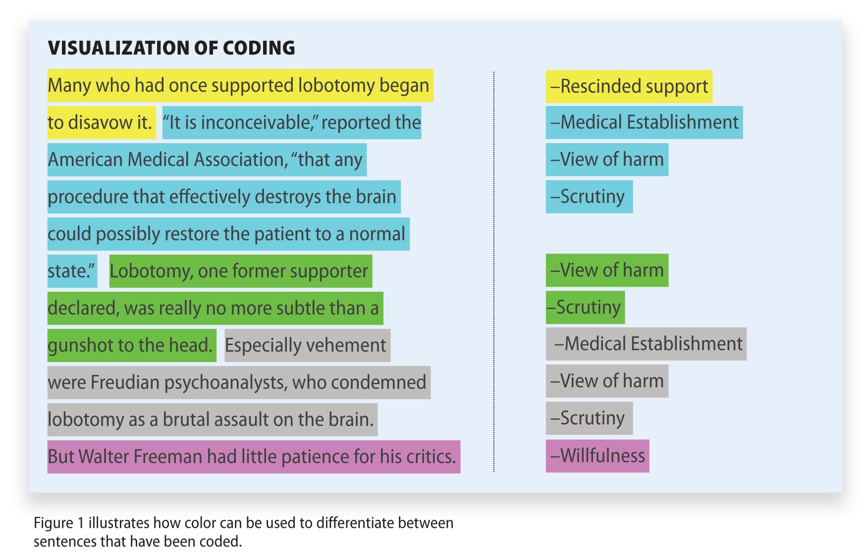
Qualitative Data Coding

What Is a Focus Group?

Cross-Cultural Research Methodology In Psychology

What Is Internal Validity In Research?

Research Methodology , Statistics
What Is Face Validity In Research? Importance & How To Measure

Criterion Validity: Definition & Examples

EP717 Module 5 - Epidemiologic Study Designs – Part 2:
Case-control studies.
- Page:
- 1
- | 2
- | 3
- | 4
- | 5
- | 6
- | 7

When is it Desirable to Conduct a Case-Control Study?
Advantages and disadvantages of case-control studies.

Case-control studies provide a method that avoids many of the limitations of cohort studies. Case-control studies are advantageous under the following circumstances:
- When exposure data are expensive or difficult to obtain, e.g., assessing pesticide levels in blood or other medical tests
- When the disease has a long induction and/or latent period, e.g., cancer, dementia. With a case-control study one does not have to wait for disease to occur,
- When the outcome (disease) is rare. (A cohort study would require too large a sample size, e.g., when studying rare parasitic diseases.)
- When the study population is dynamic and it is difficult to maintain follow up, e.g., a homeless population
- When little is known about a disease, case-control studie can evaluate multiple exposures, e.g., in the early studies of AIDS.
return to top | previous page | next page
Content ©2021. All Rights Reserved. Date last modified: April 21, 2021. Wayne W. LaMorte, MD, PhD, MPH
- Open access
- Published: 04 May 2024
Impacts of heat exposure in utero on long-term health and social outcomes: a systematic review
- Nicholas Brink 1 ,
- Darshnika P. Lakhoo 1 ,
- Ijeoma Solarin 1 ,
- Gloria Maimela 1 ,
- Peter von Dadelszen 2 ,
- Shane Norris 3 ,
- Matthew F. Chersich 1 &
Climate and Heat-Health Study Group
BMC Pregnancy and Childbirth volume 24 , Article number: 344 ( 2024 ) Cite this article
512 Accesses
8 Altmetric
Metrics details
Climate change, particularly global warming, is amongst the greatest threats to human health. While short-term effects of heat exposure in pregnancy, such as preterm birth, are well documented, long-term effects have received less attention. This review aims to systematically assess evidence on the long-term impacts on the foetus of heat exposure in utero.
A search was conducted in August 2019 and updated in April 2023 in MEDLINE(PubMed). We included studies on the relationship of environmental heat exposure during pregnancy and any long-term outcomes. Risk of bias was assessed using tools developed by the Joanna-Briggs Institute, and the evidence was appraised using the GRADE approach. Synthesis without Meta-Analysis (SWiM) guidelines were used.
Eighteen thousand six hundred twenty one records were screened, with 29 studies included across six outcome groups. Studies were mostly conducted in high-income countries ( n = 16/25), in cooler climates. All studies were observational, with 17 cohort, 5 case-control and 8 cross-sectional studies. The timeline of the data is from 1913 to 2019, and individuals ranged in age from neonates to adults, and the elderly. Increasing heat exposure during pregnancy was associated with decreased earnings and lower educational attainment ( n = 4/6), as well as worsened cardiovascular ( n = 3/6), respiratory ( n = 3/3), psychiatric ( n = 7/12) and anthropometric ( n = 2/2) outcomes, possibly culminating in increased overall mortality ( n = 2/3). The effect on female infants was greater than on males in 8 of 9 studies differentiating by sex. The quality of evidence was low in respiratory and longevity outcome groups to very low in all others.
Conclusions
Increasing heat exposure was associated with a multitude of detrimental outcomes across diverse body systems. The biological pathways involved are yet to be elucidated, but could include epigenetic and developmental perturbations, through interactions with the placenta and inflammation. This highlights the need for further research into the long-term effects of heat exposure, biological pathways, and possible adaptation strategies in studies, particularly in neglected regions. Heat exposure in-utero has the potential to compound existing health and social inequalities. Poor study design of the included studies constrains the conclusions of this review, with heterogenous exposure measures and outcomes rendering comparisons across contexts/studies difficult.
Trial Registration
PROSPERO CRD 42019140136.
Peer Review reports
Introduction
Climate change is one of the most significant threats to human health [ 1 ], characterized by an increase in global temperatures amongst other environmental changes. Global temperatures have increased by approximately 1·2 °C, and are projected to increase beyond a critical threshold of 1·5 °C in the next 5–10 years [ 2 ]. Increasingly, heat exposure is being linked with a multitude of short- and long-term health effects in vulnerable populations, including children [ 3 ], the elderly, and pregnant women [ 4 ]. The effect on pregnant women extends to the health of the foetus, with significant detrimental effects associated with heat exposure including preterm birth, stillbirth, and decreased birth weight [ 5 ]. Impacts of heat exposure are increasingly important in populations in resource-constrained settings, where heat adaptation measures such as active (air-conditioning) and passive cooling (water, green and blue spaces) are limited, and often inaccessible [ 6 ]. These populations are often found in some of the hottest climates and in areas whose contribution to global warming is negligible, thus compounding inequities [ 7 ]. In addition, research in this field is biased towards Europe, North America and Asia and is profoundly underrepresented in Africa and South America [ 8 ]. Understanding the scope and distribution of research conducted is key to guiding future research, including biological studies to explore possible mechanisms, and interventional studies to alleviate any observed negative effects. Multiple previous systematic reviews have explored the short-term impacts of heat on the foetus [ 3 , 5 , 9 ] but only one has explored the long-term impacts of heat exposure on mental health [ 10 ]. The in-utero environment has long been considered important in the long-term health and wellbeing of individuals [ 11 , 12 ], although it has been challenging to delineate specific causal pathways. This study aims to systematically review the literature on the long-term effects of heat exposure in-utero on the foetus, and explore possible casual pathways.
Materials and methods
This review forms part of a larger systematic mapping survey of the effect of heat exposure, and adaptation interventions on health (PROSPERO CRD 42019140136) [ 13 ]. The initial literature search was conducted in September 2018, where the authors searched MEDLINE (PubMed), Science Citation Index Expanded, Social Sciences Citation Index, and Arts and Humanities Citation Index using a validated search strategy (Supplementary Text 1 ). This search was updated in April 2023 through a MEDLINE search, as all previous articles were located in this database. Screening of titles and abstracts was done independently in duplicate, with any differences reconciled by MFC, with subsequent updates conducted by NB and DL. The authors only included studies on humans, published in Chinese, English, German, or Italian. Studies on heat exposure from artificial and endogenous sources were excluded, and only exogenous, weather-related heat exposure during pregnancy was included. All study designs were eligible except modelling studies and systematic reviews. No date restrictions were applied. EPPI-Reviewer software [ 14 ] provided a platform for screening, reviewing of full text articles, and for data extraction. No additional information was requested or provided by the authors. Long-term effects were defined as any outcomes that were not apparent at birth.
Articles meeting the eligibility criteria were extracted in duplicate after the initial search and then by a single reviewer in the subsequent update (NB/DL). Data were extracted to include characteristics outlined in Supplementary file 1 .
This systematic review was conducted according to the Systematic Review without Meta-Analysis (SWiM) guidelines, broadly based on PRISMA [ 15 ], as the outcomes, statistical techniques, and heat exposure measurements were heterogenous, rendering a meta-analysis untenable. Outcomes were grouped clinically, reviewed for the magnitude and direction of effect, and their statistical significance, and included negative or null findings when reported on. A text-based summary of these findings was made. ‘Vote-counting’ was utilized to summarise direction of effect findings. Analysis was conducted on the geographical areas, climate zones [ 16 ], mean annual temperature and socioeconomic classification of the country where the studies were conducted. Furthermore, an attempt was made to identify at-risk population sub-groups.
The principal investigator assessed each study for a risk of bias using the tools developed by the Joanna-Briggs Institute (JBI) [ 17 ] (Supplementary file 1 ). Each study was classified as high or low risk of bias. Studies that did not score ‘yes’ on two or more applicable parameters were classified as high risk of bias [ 5 ]. Due to the limited research in this field, no studies were excluded based on risk of bias. The certainty of the evidence was assessed using the GRADE approach, with the body of evidence assessed on a scale of certainty: very low, low, moderate and high [ 18 ]. Due to the heterogeneity of outcomes, and the reporting thereof, assessment of publication bias was not possible.
The funder of the study had no role in study design, data collection, analysis, interpretation, or writing of the report.
The updated search identified 18 621 non-duplicate records, and after screening 229 full-text articles were reviewed for inclusion, with a total of 29 studies included in the final analysis (Fig. 1 : flow chart). The included studies were conducted in 25 countries across six continents, including six Low-Income Countries (LIC), two Lower-Middle Income Countries (LMIC), one Upper-Middle Income Country (UMIC) and 16 High Income Countries (HIC) [ 19 ]. They included 25 Köppen-Geiger climate zones [ 16 ], and mean annual temperatures ranging from 2.1 °C in Norway to 30.0 °C in Burkina Faso [ 20 ] (Figs. 2 and 3 ). All studies were observational, with 17 cohort, five case-control and eight cross-sectional studies. The timeline of the data is from 1913 to 2019, and individuals included ranged in age from neonates to adults, and the elderly. The studies were grouped by outcomes as follows: behavioural, educational and socioeconomic ( n = 6), cardiovascular disease ( n = 6), respiratory disease ( n = 3), growth and anthropometry ( n = 2), mental health ( n = 12) and longevity and mortality ( n = 3). The measures of heat exposure were variable, with minimum, mean, maximum, and apparent temperature being utilized, as well as temperature variability, heat wave days and discreet shocks (number of times exposure exceeded a specific threshold). The majority of studies measured heat using mean temperature ( n = 27/29). In addition, the statistical comparison was diverse, with some studies making a continuous linear comparison by degree Celsius, while others compared heat exposure by quartiles, amongst other categorical comparisons. Furthermore, heat exposure by any definition was not reported over the same timeframes, with some studies including variable periods before birth, during pregnancy and at birth in their analysis. Levels of temporal resolution of heat exposure were also diverse, ranging from monthly effects to effects observed over the entire gestational period, or year of birth. In addition, differing use of heat adaptation mechanisms was not uniformly described and adjusted for. Various confounders were adjusted for, and although not uniform, these were generally inadequate. The effect on female infants was greater than on male infants in eight of nine studies differentiating by sex, with increased effects on marginalised groups (African-Americans) in one further study. Overall, the quality of the evidence, as assessed by the GRADE approach, was low in respiratory and longevity outcome groups to very low in all other groups, primarily as a result of their observational nature and high risk of bias, due to insufficient consideration of confounders, and inadequate measures of heat exposure.
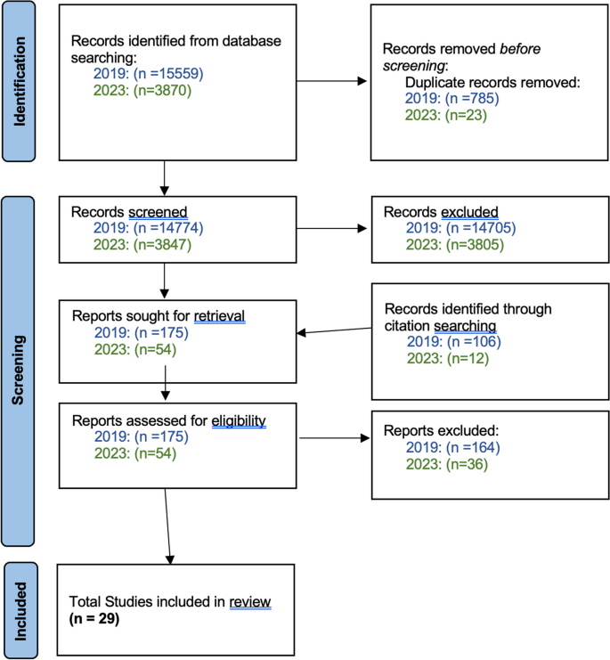
PRISMA flow diagram
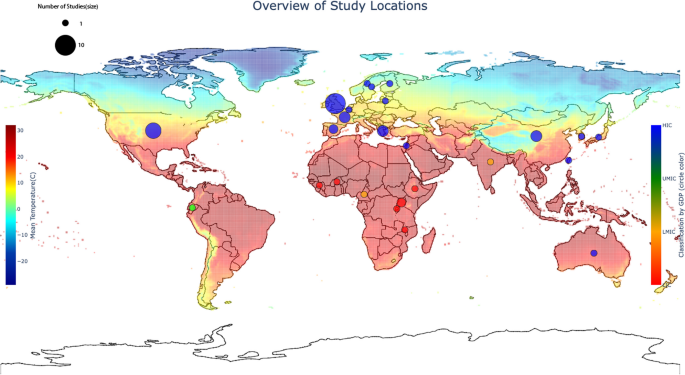
Map showing countries where studies were conducted relative to mean annual temperature [ 21 ]
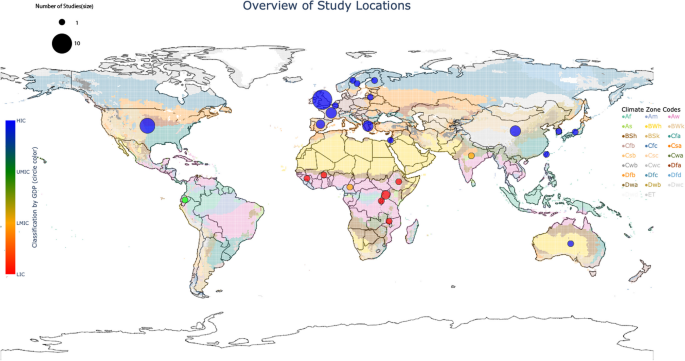
Map showing countries where studies were conducted relative to climate zones [ 16 ]
A total of six studies reported on behaviour, educational and socioeconomic outcomes, which were detrimentally affected by increases in heat exposure (Fig. 4 ; Table 1 ), although the quality of the evidence was very low . End-points were not uniform, but included earnings, completion of secondary school or higher education, number of years of schooling, and gamified cooperation-rates in a public-goods game (where test scores represent achieving maximal public benefit in hypothetical situations).
Two large studies reported a detrimental effect of heat exposure on adult income, with the greatest effect noted in first trimester exposure. These studies noted a reduction in earnings of up to 1·2% per 1 °C increase in temperature, with greater effects in females [ 22 ], and a decrease of $55.735 (standard error(SE): 15·425, P < 0·01) annual earnings at 29–31 years old, per day exposure > 32 °C [ 26 ]. Two studies reported worse educational outcomes, with the greatest effect noted in the second trimester [ 23 ]. Rates of completing secondary education were found to be reduced by 0·2% per 1 °C increase in temperature ( P = 0·05) [ 22 ], illiteracy was increased by 0·18% (SE=(0·0009); P < 0·05) and mean years of schooling was lowered by 0.02 (SE=(0·009) P = 0·07) [ 23 ]. Two studies reported a beneficial effect of heat exposure on educational outcomes, although both studies suffered from significant methodological flaws, and effects were < 0·01% when effect estimates were noted [ 24 , 27 ]. One small study reported lower cooperation rates by 20% ( P < 0·01) in a public-goods game, with lower predicted public wellbeing [ 25 ].
The studies generally exhibited a dose-response effect with evidence for a critical threshold of effect of 28 °C in one study [ 22 ]. All studies were at a high risk of bias.
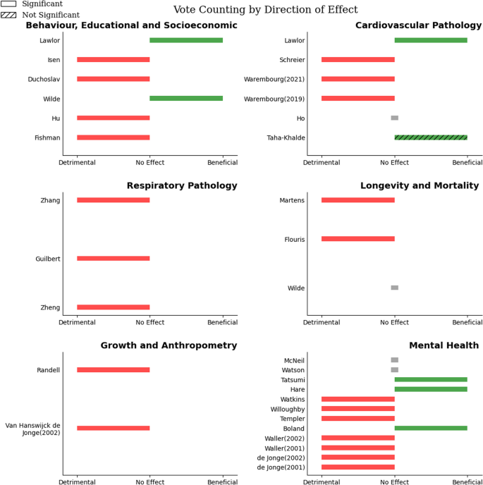
Figure showing vote counting across all outcome groups. No Effect = No direction of effect noted in study
Six studies reported on cardiovascular pathology and risk factors thereof, which were detrimentally affected by increased exposure to heat (Fig. 4 ; Table 2 ), although measures and surrogates of this outcome were heterogenous. The quality of the evidence was very low, and the sample sizes were small. Outcomes included blood pressure, a composite cardiovascular disease indicator, and specific cardiovascular disease risk factors such as diabetes mellitus (type I), insulin resistance, waste circumference, and triglyceride levels.
Three studies found a detrimental effect of heat exposure on hypertension rates, and increased blood pressure [ 31 ], with a maximum of 1·6 mm Hg increase noted per interquartile range (IQR) increase (95% Confidence interval (CI) = 0·2, 2·9, P = 0·024) in children [ 30 ], with increased effects on women in the largest study ( N = 11,237) [ 32 ]. Another study found increasing heat exposure at conception was detrimentally associated with an increase in coronary heart disease ( P = 0·08) [ 32 ], although one of the smaller studies ( N = 4286) found a beneficial effect of heat exposure at birth on diverse cardiovascular outcomes, including coronary heart disease ( P = 0·03 for trend), triglyceride levels ( P = 0·06 for trend) and insulin resistance ( P = 0·04 for trend) [ 27 ]. One study found lower odds of type I diabetes mellitus with increasing heat exposure, with odds ratio (OR) = 0·73 (95%CI = 0·48, 1·09, P -value not stated) [ 28 ]. Another study did not detect statistically significant relationships between heat exposure and hypertension or a composite cardiovascular disease indicator, but did not provide effect estimates [ 29 ]. Five studies were at a high risk of bias [ 27 , 29 , 30 , 31 , 32 ], with only one case-control study at a low risk of bias [ 28 ].
Respiratory pathology was reported by three studies, assessing different outcomes. Outcomes were detrimentally associated with increasing heat (Fig. 4 ; Table 3 ), however the quality of the evidence was low . The outcomes were primarily measured in infants and children, with no studies on adult outcomes. The largest study ( N = 1681) found that increasing heat exposure increased the odds of having childhood asthma [ 33 ], and another small study ( N = 343) noted worsened lung function with increasing heat exposure [ 34 ].
An additional study noted increased odds of childhood pneumonia with increasing diurnal temperature variation (DTV) in pregnancy, with a maximum OR = 1·85 (95%CI = 1·24, 2·76) in the third trimester [ 35 ].
Exposure in the third trimester had the greatest effect across all three studies [ 33 , 34 , 35 ]. Females showed an increased susceptibility to heat exposure’s effects on lung function, but males were more susceptible to heat’s effect on childhood pneumonia. There was a critical threshold noted in the asthma study of 24·6 °C, with a dose-response effect. The asthma study was assessed as low risk of bias, however the other studies were at high risk.
Growth and anthropometry was reported on by two studies, with differing outcomes, although in both, heat exposure was associated with detrimental, although heterogenous, outcomes (Fig. 4 ; Table 4 ). The overall quality of the evidence was very low . One study found a positive association with heat exposure and increased body mass index (BMI), r = 0·22 ( P < 0·05) in the third trimester with greater effects noted in females and in African-Americans [ 36 ]. Another large study ( N = 23 026) found increased odds of stunting (OR = 1·28, 95%CI = not stated, p < 0·001) with a negative correlation with height noted ( r =-0·083 P < 0·01) [ 37 ]. Effects were greatest in the first and third trimester. Both studies were at a high risk of bias.
Mental health was reported on by 12 studies. Increasing heat exposure generally had a detrimental association with mental health outcomes (Fig. 4 ; Table 5 ), although these were heterogenous. The overall quality of the evidence was very low . Five studies reported on schizophrenia rates, with only one study showing a strongly positive association of heat exposure at conception with schizophrenia rates ( r = 0·50, p < 0·025) [ 38 ]. Another study noted the same effect with increasing heat in the summer before birth, however this was not statistically significant [ 39 ]. The third study reported no association of this outcome [ 40 ], with another small study ( N = 2985) showing a negative correlation with temperatures at birth, without reporting on heat exposure during other periods of gestation [ 41 ]. The fifth study failed to report direction of effect, but noted non-significant findings [ 42 ]. Six studies reported on eating disorders, with all six showing a detrimental effect with increasing heat exposure. Of the three studies on clinical anorexia nervosa, one reported increasing rates of anorexia nervosa compared to other eating disorders (χ²= 4·48, P = 0·017) [ 43 ], another reported increasing rates of a restrictive-subtype (χ²= 3·18, P = 0·04) as well as reporting worse assessments of restrictive behaviours [ 44 ], which was supported by a third study in a different setting [ 45 ]. Three studies examined non-clinical settings, with some inconsistent effects. The first study showed a weak positive association with heat exposure, and drive for thinness (Spearman’s ⍴ = 0·46, P < 0·05) and bulimia scores (Spearman’s ⍴ = 0·25, P < 0·05) [ 46 ], which was supported by a replication study [ 47 ], and one other study [ 48 ]. The most significant and consistent effects noted in the third trimester, at birth, and in females [ 47 , 48 ]. One study reported a beneficial effect of increased temperatures in the first trimester on rates of depression, however no other directions of effect were noted for other periods of exposure [ 49 ]. These studies were at a high risk of bias.
Increasing heat exposure had a detrimental effect on longevity and mortality across various outcomes (Fig. 4 ; Table 6 ), although despite large sample sizes, the quality of the evidence was low . One study found a negative correlation of heat exposure with longevity ( r =-0·667, P < 0·001), with a greater effect on females [ 50 ]. A second study showed a detrimental effect on telomere length, as a predicter of longevity, with the greatest effect towards the end of gestation (3·29% shorter TL, 95%CI = − 4·67, − 1·88, per 1 C increase above 95th centile) [ 51 ]. Conversely, a third study noted no correlation with mortality [ 24 ]. All but the study on telomere length [ 51 ] were at a high risk of bias.
This study establishes significant patterns of effects amongst the outcomes reviewed, with increasing heat exposure being associated with an overall detrimental effect on multiple, diverse, long-term outcomes. These effects are likely to increase with rising temperatures, however modelling this is beyond the scope of this review.
The most notable detrimental outcomes are related to neurodevelopmental pathways, with behavioural, educational, socioeconomic and mental health outcomes consistently associated with increasing heat exposure, in addition to having the greatest body of literature to support this. Importantly, other systems such as the respiratory and cardiovascular systems also suggest harmful effects of heat exposure, culminating in detrimental associations with longevity and mortality. Some studies illustrated a possible beneficial effect in some disease-processes, such as coronary heart disease and depression showing the potential for shifting disease profiles with rising temperatures.
The detrimental effects of heat exposure became more significant with increasing temperatures, with many studies describing increasing effects beyond critical thresholds which, although varied across studies, suggest that there is a limit of heat adaption strategies, both biological and behavioural [ 52 , 53 ].
In addition, the effect of increasing heat exposure was associated with worse outcomes in already marginalised communities, such as women [ 22 , 32 , 34 , 36 , 44 , 47 , 48 , 50 ] and certain ethnic groups (African-Americans) [ 46 ]. The reasons for sub-population vulnerabilities are unclear and likely complex. In the case of female foetuses being more susceptible to changes in the in-utero environment, it is possible that there is a ‘survivorship bias’. This would occur if women with harmful exposure lose male infants during pregnancy at a higher rate, and thus the surviving female infants appear more at risk. However, despite an increased risk of early pregnancy loss, there are no studies that have assessed this differential vulnerability. This still has the effect of potentially increasing the burden of disease on an already marginalised group.
In the case of certain population groups being more at risk, it is likely that both physiological differences in vulnerability as well as socio-economic effect-modifiers exist to explain these differences, however, the included literature lacks sufficient evidence to assess this. The vulnerabilities of different populations to the long-term effects of heat exposure in-utero likely contributes to the unequal impacts of climate change that have already been established [ 54 ], and will be an important contributor to inequality with future increases in temperature. Further research in this area is critical to inform targeted redistributive interventions.
Although the associations may be clear, establishing causality is fraught with difficulty, with no consensus on an infallible approach [ 55 , 56 , 57 ]. However, it is prudent to highlight supporting evidence in this review.
The hypothesis that the in-utero environment had significant long-term impacts on the foetus was first suggested by Barker, in the context of maternal nutrition and cardiovascular disease [ 11 ]. Further studies supported this hypothesis, and expanded on the effects the in-utero environment has on the foetus and its long-term wellbeing [ 58 ]. Long-term heat exposure may also be associated with changes in nutritional availability [ 11 ], and is likely one of many complex but important environmental exposures in-utero.
Maternal comorbidities, associated with increasing heat exposure such as hypertensive disorders of pregnancy and gestational diabetes mellitus, are known to negatively affect the foetus in the long-term [ 59 , 60 ]. These comorbidities may be part of the long-term pathogenicity of heat exposure, through short-term exposure-outcome pathways. Placental dysfunction is central to the pathology of pre-eclampsia, and is a significant cause for foetal pathology [ 61 , 62 ]. The placenta is not auto-regulated and is therefore acutely affected by changes to blood volume, heart rate and blood pressure, culminating in cardiac output as it is delivered to the placenta as an end-organ with resultant negative effects on the foetus [ 63 ]. Heat-acclimatisation mechanisms are hypothesized to affect this delicate balance [ 52 , 64 ], with observational studies supporting this [ 64 ]. It has been suggested that heat exposure’s increase in inflammation is a possible causative mechanism for pre-term birth [ 5 , 52 ], but inflammation has numerous additional effects on the immune system and could prove an insult to the mother and developing foetus [ 62 , 65 ]. These effects may only manifest in the long-term.
Heat was one of the earliest described teratogens [ 66 ], with significant effects on neurodevelopment noted in animal models in keeping with the observed associations of this review [ 67 ]. Biological organisms are extremely dependent on heat as a trigger for various processes. Plants and animals undergo significant change in response to the seasons, which are often guided by fluctuations in temperature. These changes are often mediated by epigenetic mechanisms, allowing the modification and modulation of gene expression [ 68 , 69 ].
Thus, from an evolutionary perspective, DNA, is sensitive to changes in temperature. The mechanism of this sensitivity has been shown to be primarily epigenetic in nature [ 69 ]. Increasing heat results in modifications to histone deacetylation and DNA methylation [ 69 ]. This is required to provide fast-acting adaptions to acute stressors, but can have long-term effects too [ 70 ]. Thus, it is likely that humans are sensitive to changes in temperature, which can alter epigenetic modifications, and thus our exposome. This sensitivity, may have provided a survival benefit in times of increasing heat, or it may simply be a vestigial function which provides no survival benefit, and may in fact have detrimental effects [ 71 ]. Epigenetic changes have been shown to have significant effects on metabolic diseases and risk profiles, and an in-depth review is provided by Wu et al. [ 72 ]. The exact processes and genes involved would be an area requiring further research, where similar research exists on the effects of nutrition on exact epigenetic pathways [ 73 ]. An important pattern requiring further research involves the effect heat may have on neurodevelopment [ 67 , 74 ]. The above pathways provide additional mechanisms for the long-term lag between exposure-outcome pathways. In addition, acute heat exposure at the time of birth has been associated with various possibly pathogenic mechanisms such as preterm birth [ 5 ], low APGAR scores [ 75 ] and foetal distress [ 76 ], as well as a possible effect on the maternal microbiome and the seeding thereof to the neonate [ 10 , 64 , 77 , 78 ]. These effects, can all provide plausible causes for the long-term outcomes observed through short-term insults. The interplay of these, and additional factors is highlighted in Fig. 5 [ 79 ]. Importantly, the periods of vulnerability are likely different for these various pathways, but specific outcomes may have multiple periods of vulnerability through different pathways.
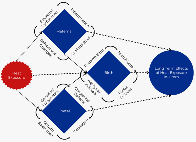
Causal pathways
The outcomes associated with increasing heat exposure highlight the health, social, and economic cost of global warming, establishing current estimates and future predictions for this are beyond the scope of this research but would provide a valuable area for future research. This would entail estimating disease-burden due to climate change through attribution studies. Traditional health impact studies conflate adverse outcomes from natural variations in climate (‘noise’) with adverse outcomes from anthropogenic climate change. However, not every climate-related adverse outcome is the result of anthropogenic climate change, and these effects are likely different in vulnerable populations. This highlights the benefit of studying and implementing effective heat adaptation strategies in areas where the greatest effect is likely to be observed, and where the greatest impacts in lessening the economic and human impact of global warming are possible [ 80 , 81 ].
Limitations
The difficulty in assessing the data is compounded by the heterogenous measures of heat exposure. No studies used widely accepted heat exposure indices that consider important environmental modifying factors like humidity and windspeed [ 82 , 83 ]. In addition, effect modifiers, heat acclimatisation and adaptation strategies were seldom considered [ 84 , 85 , 86 ]. It may be prudent for future studies to consider the measure of ionizing radiation exposure as an analogous environmental exposure, where different measures exist for the intensity, total quantity (a function of duration of exposure) and biologically-adjusted quantity absorbed [ 87 ]. Differing time-periods of exposure made it difficult to evaluate specific periods of sensitivity, which are likely different for various outcomes, depending on critical periods of development.
Despite consistency across different contexts in this review, the analysis of the distribution of the included studies highlights the unequal weight of studies towards relatively cooler climates, in regions with higher socioeconomic levels and likely greater heat adaptation uptake, and must therefore be interpreted in this context. It is possible that myriad factors that differ geographically, including physiological and socio-economic differences, will influence the effects of heat, and thus there is likely no underlying universal truth to associations and effect estimates.
Quantifying, describing and comparing the effect size across studies was rendered more difficult due to heterogenous statistical analyses.
Although some studies adjusted for possible confounding variables, not all reported on this, with the effects of seasonal, foetal, and maternal biological factors that may not lie on the causal pathway seldom considered [ 3 , 5 , 9 , 88 , 89 , 90 , 91 , 92 ].
Data extraction and assessment of risk of bias was not uniformly undertaken in duplicate due to resource constraints, which may predispose to extraction errors or bias. The high risk of bias of included studies, limits the utility of the overall assessment of effects and suggestions for further action. In addition, publication bias is likely skewing the results towards statistically significant detrimental results, with studies with smaller sample sizes not necessarily showing wider distribution of findings as would be expected.
Climate change, and in particular, global warming, is a significant emerging global public health threat, with far reaching, and disproportionate effects on the most vulnerable populations. The effects of increasing heat exposure in utero are associated with, and possibly causal in, wide-ranging long-term impacts on socioeconomic and health outcomes with a significant cost associated with increasing global temperatures. This association is as a result of a complex interplay of factors, including through direct and indirect effects on the mother and foetus. Further research is urgently required to elicit biological pathways, and targets for intervention as well as predicting future disease-burden and economic impacts through attribution studies.
Availability of data and materials
This study was a review of publicly available information data, with references to data sources made in the reference list.
Abbreviations
Apparent Temperature
Body Mass Index
Blood Pressure
Coronary Heart Disease
Confidence Interval
Diastolic Blood Pressure
Eating Disorder Inventory
Functional Residual Capacity
Interquartile Range
Non-Significant
Respiratory Rate
Systolic Blood Pressure
Standard Error
Telomere Length
World Health Organisation
Atwoli L, A HB, Benfield T, et al. Call for emergency action to limit global temperature increases, restore biodiversity and protect health. BMJ Lead. 2022;6(1):1–3.
Article PubMed Google Scholar
Organization WM, World Meteorological O. WMO global annual to decadal climate update (Target years: 2023–2027). Geneva: WMO; 2023. p. 24.
Book Google Scholar
Lakhoo DP, Blake HA, Chersich MF, Nakstad B, Kovats S. The effect of high and low ambient temperature on infant health: a systematic review. Int J Environ Res Public Health. 2022;19(15):9109.
Article PubMed PubMed Central Google Scholar
Cissé G, McLeman R, Adams H, et al. Health, wellbeing and the changing structure of communities. In: Pörtner HO, Roberts DC, Tignor MMB, Poloczanska ES, Mintenbeck K, Alegría A, et al. editors. Climate change 2022: impacts, adaptation and vulnerability contribution of working group II to the sixth assessment report of the intergovernmental panel on climate change. Cambridge: Cambridge University Press; 2022.
Chersich MF, Pham MD, Areal A, et al. Associations between high temperatures in pregnancy and risk of preterm birth, low birth weight, and stillbirths: systematic review and meta-analysis. BMJ. 2020;371:m3811.
Birkmann J, Liwenga E, Pandey R, et al. Poverty, livelihoods and sustainable development. In: Pörtner HO, Roberts DC, Tignor MMB, Poloczanska ES, Mintenbeck K, Alegría A, et al. editors. Climate change 2022: impacts, adaptation and vulnerability contribution of working group II to the sixth assessment report of the intergovernmental panel on climate change. Cambridge: Cambridge University Press; 2022.
Watts N, Amann M, Ayeb-Karlsson S, et al. The Lancet countdown on health and climate change: from 25 years of inaction to a global transformation for public health. Lancet. 2018;391(10120):581–630.
Campbell-Lendrum D, Manga L, Bagayoko M, Sommerfeld J. Climate change and vector-borne diseases: what are the implications for public health research and policy? Philos Trans R Soc Lond B Biol Sci. 2015;370(1665):20130552.
Haghighi MM, Wright CY, Ayer J, et al. Impacts of high environmental temperatures on congenital anomalies: a systematic review. Int J Environ Res Public Health. 2021;18(9):4910.
Puthota J, Alatorre A, Walsh S, Clemente JC, Malaspina D, Spicer J. Prenatal ambient temperature and risk for schizophrenia. Schizophr Res. 2022;247:67–83.
Barker DJ. Intrauterine programming of coronary heart disease and stroke. Acta Paediatr Suppl. 1997;423:178–82; discussion 83.
Article CAS PubMed Google Scholar
Hales CN, Barker DJ. The thrifty phenotype hypothesis. Br Med Bull. 2001;60:5–20.
Manyuchi A, Dhana A, Areal A, et al. Title: systematic review to quantify the impacts of heat on health, and to assess the effectiveness of interventions to reduce these impacts. PROSPERO: International prospective register of systematic reviews. 2019, 42019140136. Available from: https://www.crd.york.ac.uk/PROSPEROFILES/118113_PROTOCOL_20181129.pdf .
Thomas J, Brunton J, Graziosi S. EPPI-Reviewer 4.0: software for research synthesis. EPPI Centre Software. London: Social Science Research Unit, Institute of Education, University of London; 2010.
Campbell M, McKenzie JE, Sowden A, et al. Synthesis without meta-analysis (SWiM) in systematic reviews: reporting guideline. BMJ. 2020;368:l6890.
Beck HE, Zimmermann NE, McVicar TR, Vergopolan N, Berg A, Wood EF. Present and future Köppen-Geiger climate classification maps at 1-km resolution. Sci Data. 2018;5(1):180214.
Barker TH, Stone JC, Sears K, et al. Revising the JBI quantitative critical appraisal tools to improve their applicability: an overview of methods and the development process. JBI Evid Synth. 2023;21(3):478.
Higgins JPT, Thomas J, Chandler J, Cumpston M, Li T, Page MJ, Welch VA (editors). Cochrane Handbook for Systematic Reviews of Interventions version 6.4 (updated August 2023). Cochrane, 2023. Available from www.training.cochrane.org/handbook .
World Bank Country and Lending Groups – World Bank Data Help Desk. Retrieved: June 01 2023. Available from: https://datahelpdesk.worldbank.org/knowledgebase/articles/906519-world-bank-country-and-lending-groups-files/112/906519-world-bank-country-and-lending-groups.html .
Home | Climate change knowledge portal. Retrieved: June 01 2023. Available from: https://climateknowledgeportal.worldbank.org/files/26/climateknowledgeportal.worldbank.org.html .
Harris I, Osborn TJ, Jones P, Lister D. Version 4 of the CRU TS monthly high-resolution gridded multivariate climate dataset. Sci Data. 2020;7(1):109.
Fishman R, Carrillo P, Russ J. Long-term impacts of exposure to high temperatures on human capital and economic productivity. J Environ Econ Manag. 2019;93:221–38.
Article Google Scholar
Hu Z, Li T. Too hot to handle: the effects of high temperatures during pregnancy on adult welfare outcomes. J Environ Econ Manag. 2019;94:236–53.
Wilde J, Apouey BH, Jung T. The effect of ambient temperature shocks during conception and early pregnancy on later life outcomes. Eur Econ Rev. 2017;97:87–107.
Duchoslav J. Prenatal temperature shocks reduce cooperation: evidence from public goods games in Uganda. Front Behav Neurosci. 2017;11: 249.
Isen A, Rossin-Slater M, Walker R. Relationship between season of birth, temperature exposure, and later life wellbeing. Proc Natl Acad Sci U S A. 2017;114(51):13447–52.
Article CAS PubMed PubMed Central Google Scholar
Lawlor DA, Davey Smith G, Mitchell R, Ebrahim S. Temperature at birth, coronary heart disease, and insulin resistance: cross sectional analyses of the British women’s heart and health study. Heart. 2004;90(4):381–8.
Taha-Khalde A, Haim A, Karakis I, et al. Air pollution and meteorological conditions during gestation and type 1 diabetes in offspring. Environ Int. 2021;154: 106546.
Ho JY. Early-life environmental exposures and height, hypertension, and cardiovascular risk factors among older adults in India. Biodemography Soc Biol. 2015;61(2):121–46.
Warembourg C, Maitre L, Tamayo-Uria I, et al. Early-life environmental exposures and blood pressure in children. J Am Coll Cardiol. 2019;74(10):1317–28.
Warembourg C, Nieuwenhuijsen M, Ballester F, et al. Urban environment during early-life and blood pressure in young children. Environ Int. 2021;146: 106174.
Schreier N, Moltchanova E, Forsen T, Kajantie E, Eriksson JG. Seasonality and ambient temperature at time of conception in term-born individuals - influences on cardiovascular disease and obesity in adult life. Int J Circumpolar Health. 2013;72: 21466.
Zhang J, Bai S, Lin S, et al. Maternal apparent temperature during pregnancy on the risk of offspring asthma and wheezing: effect, critical window, and modifiers. Environ Sci Pollut Res Int. 2023;30(22):62924–37.
Guilbert A, Hough I, Seyve E, et al. Association of prenatal and postnatal exposures to warm or cold air temperatures with lung function in young infants. JAMA Netw Open. 2023;6(3):e233376.
Zheng X, Kuang J, Lu C, et al. Preconceptional and prenatal exposure to diurnal temperature variation increases the risk of childhood pneumonia. BMC Pediatr. 2021;21(1):192.
van Hanswijck de Jonge L, Stettler N, Kumanyika S, Stoa Birketvedt G, Waller G. Environmental temperature during gestation and body mass index in adolescence: new etiologic clues? Int J Obes Relat Metab Disord. 2002;26(6):765–9.
Randell H, Gray C, Grace K. Stunted from the start: early life weather conditions and child undernutrition in Ethiopia. Soc Sci Med. 2020;261: 113234.
Templer DI, Austin RK. Confirmation of relationship between temperature and the conception and birth of schizophrenics. J Orthomolecular Psychiatr. 1980;9(3):220-2.
Hare E, Moran P. A relation between seasonal temperature and the birth rate of schizophrenic patients. Acta Psychiatr Scand. 1981;63(4):396–405.
Watson CG, Kucala T, Tilleskjor C, Jacobs L. Schizophrenic birth seasonality in relation to the incidence of infectious diseases and temperature extremes. Arch Gen Psychiatry. 1984;41(1):85–90.
Tatsumi M, Sasaki T, Iwanami A, Kosuga A, Tanabe Y, Kamijima K. Season of birth in Japanese patients with schizophrenia. Schizophr Res. 2002;54(3):213–8.
McNeil T, Dalén P, Dzierzykray-Rogalska M, Kaij L. Birthrates of schizophrenics following relatively warm versus relatively cool summers. Arch Psychiat Nervenkr. 1975;221(1):1–10.
Watkins B, Willoughby K, Waller G, Serpell L, Lask B. Pattern of birth in anorexia nervosa I: early-onset cases in the United Kingdom. Int J Eat Disord. 2002;32(1):11–7.
Waller G, Watkins B, Potterton C, et al. Pattern of birth in adults with anorexia nervosa. J Nerv Ment Dis. 2002;190(11):752.
Willoughby K, Watkins B, Beumont P, Maguire S, Lask B, Waller G. Pattern of birth in anorexia nervosa. II: a comparison of early-onset cases in the southern and northern hemispheres. Int J Eat Disord. 2002;32(1):18–23.
van Hanswijck L, Meyer C, Smith K, Waller G. Environmental temperature during pregnancy and eating attitudes during teenage years: a replication and extension study. Int J Eat Disord. 2001;30(4):413–20.
van Hanswijck L, Waller G. Influence of environmental temperatures during gestation and at birth on eating characteristics in adolescence: a replication and extension study. Appetite. 2002;38(3):181–7.
Waller G, Meyer C, van Hanswijck de Jonge L. Early environmental influences on restrictive eating pathology among nonclinical females: the role of temperature at birth. Int J Eat Disord. 2001;30(2):204–8.
Boland MR, Parhi P, Li L, et al. Uncovering exposures responsible for birth season – disease effects: a global study. J Am Med Inform Assoc. 2018;25(3):275–88.
Flouris AD, Spiropoulos Y, Sakellariou GJ, Koutedakis Y. Effect of seasonal programming on fetal development and longevity: links with environmental temperature. Am J Hum Biol. 2009;21(2):214–6.
Martens DS, Plusquin M, Cox B, Nawrot TS. Early biological aging and fetal exposure to high and low ambient temperature: a birth cohort study. Environ Health Perspect. 2019;127(11):117001.
Samuels L, Nakstad B, Roos N, et al. Physiological mechanisms of the impact of heat during pregnancy and the clinical implications: review of the evidence from an expert group meeting. Int J Biometeorol. 2022;66(8):1505–13.
Ravanelli N, Casasola W, English T, Edwards KM, Jay O. Heat stress and fetal risk. Environmental limits for exercise and passive heat stress during pregnancy: a systematic review with best evidence synthesis. Br J Sports Med. 2019;53(13):799–805.
Islam SN, Winkel J. Climate change and social inequality, DESA working paper no. 152, United Nations Department of Economic and Social Affiars (2017). Available from: https://www.un.org/esa/desa/papers/2017/wp152_2017.pdf .
Rothman KJ, Greenland S. Causation and causal inference in epidemiology. Am J Public Health. 2005;95(S1):S144–150.
Fedak KM, Bernal A, Capshaw ZA, Gross S. Applying the Bradford Hill criteria in the 21st century: how data integration has changed causal inference in molecular epidemiology. Emerg Themes Epidemiol. 2015;12(1):14.
Hill AB. The environment and disease: association or causation? J R Soc Med. 2015;108(1):32–7.
Gluckman PD, Hanson MA, Cooper C, Thornburg KL. Effect of in Utero and early-life conditions on adult health and disease. N Engl J Med. 2008;359(1):61–73.
Duley L. The global impact of pre-eclampsia and eclampsia. Semin Perinatol. 2009;33(3):130–7.
Bianco ME, Kuang A, Josefson JL, et al. Hyperglycemia and adverse pregnancy outcome follow-up study: newborn anthropometrics and childhood glucose metabolism. Diabetologia. 2021;64(3):561–70.
Fisher SJ. The placental problem: linking abnormal cytotrophoblast differentiation to the maternal symptoms of preeclampsia. Reprod Biol Endocrinol. 2004;2: 53.
Steegers EAP, von Dadelszen P, Duvekot JJ, Pijnenborg R. Pre-eclampsia. Lancet. 2010;376(9741):631–44.
von Dadelszen P, Ornstein MP, Bull SB, Logan AG, Koren G, Magee LA. Fall in mean arterial pressure and fetal growth restriction in pregnancy hypertension: a meta-analysis. Lancet. 2000;355(9198):87–92.
Bonell A, Sonko B, Badjie J, et al. Environmental heat stress on maternal physiology and fetal blood flow in pregnant subsistence farmers in the Gambia, West Africa: an observational cohort study. Lancet Planet Health. 2022;6(12):e968–76.
Redline RW. Placental inflammation. Semin Neonatol. 2004;9(4):265–74.
Alsop FM. The effect of abnormal temperatures upon the developing nervous system in the chick embryos. Anat Rec. 1919;15(6):306–31.
Edwards MJ. Hyperthermia as a teratogen: a review of experimental studies and their clinical significance. Teratog Carcinog Mutagen. 1986;6(6):563–82.
Nicotra AB, Atkin OK, Bonser SP, et al. Plant phenotypic plasticity in a changing climate. Trends Plant Sci. 2010;15(12):684–92.
McCaw BA, Stevenson TJ, Lancaster LT. Epigenetic responses to temperature and climate. Integr Comp Biol. 2020;60(6):1469–80.
Horowitz M. Epigenetics and cytoprotection with heat acclimation. J Appl Physiol (1985). 2016;120(6):702–10.
Murray KO, Clanton TL, Horowitz M. Epigenetic responses to heat: from adaptation to maladaptation. Exp Physiol. 2022;107(10):1144–58.
Wu YL, Lin ZJ, Li CC, et al. Epigenetic regulation in metabolic diseases: mechanisms and advances in clinical study. Sig Transduct Target Ther. 2023;8:98.
Sookoian S, Gianotti TF, Burgueño AL, Pirola CJ. Fetal metabolic programming and epigenetic modifications: a systems biology approach. Pediatr Res. 2013;73(2):531–42.
Graham JM, Marshall J. Edwards: discoverer of maternal hyperthermia as a human teratogen. Birth Defects Res Clin Mol Teratol. 2005;73(11):857–64.
Article CAS Google Scholar
Andalón M, Azevedo JP, Rodríguez-Castelán C, Sanfelice V, Valderrama-González D. Weather shocks and health at Birth in Colombia. World Dev. 2016;82:69–82.
Cil G, Cameron TA. Potential climate change health risks from increases in heat waves: abnormal birth outcomes and adverse maternal health conditions. Risk Anal. 2017;37(11):2066–79.
Huus KE, Ley RE. Blowing hot and cold: body temperature and the microbiome. mSystems. 2021;6(5):e00707–21.
Wen C, Wei S, Zong X, Wang Y, Jin M. Microbiota-gut-brain axis and nutritional strategy under heat stress. Anim Nutr. 2021;7(4):1329–36.
ACOG Committee. Physical activity and exercise during pregnancy and the postpartum period, ACOG Committee Opinion No. 804. Obstet Gynecol. 2020;135e:88.
Google Scholar
Lee H, Calvin K, Dasgupta D, et al. Synthesis report of the IPCC Sixth Assessment Report (AR6). Geneva: IPCC; 2023. p. 35–115.
Stern N. Economics: current climate models are grossly misleading. Nature. 2016;530(7591):407–9.
McGregor GR, Vanos JK. Heat: a primer for public health researchers. Public Health. 2018;161:138–46.
Gao C, Kuklane K, Östergren P-O, Kjellstrom T. Occupational heat stress assessment and protective strategies in the context of climate change. Int J Biometeorol. 2018;62(3):359–71.
Alele F, Malau-Aduli B, Malau-Aduli A, Crowe M. Systematic review of gender differences in the epidemiology and risk factors of exertional heat illness and heat tolerance in the armed forces. BMJ Open. 2020;10(4): e031825.
Khosla R, Jani A, Perera R. Health risks of extreme heat. BMJ. 2021;375:n2438.
Spector JT, Masuda YJ, Wolff NH, Calkins M, Seixas N. Heat exposure and occupational injuries: review of the literature and implications. Curr Environ Health Rep. 2019;6(4):286–96.
Measuring Radiation. Centers for disease control and prevention. 2015. Retrieved: May 15 2023. Available from: https://www.cdc.gov/nceh/radiation/measuring.html .
Molina-Vega M, Gutiérrez-Repiso C, Muñoz-Garach A, et al. Relationship between environmental temperature and the diagnosis and treatment of gestational diabetes mellitus: an observational retrospective study. Sci Total Environ. 2020;744: 140994.
Su W-L, Lu C-L, Martini S, Hsu Y-H, Li C-Y. A population-based study on the prevalence of gestational diabetes mellitus in association with temperature in Taiwan. Sci Total Environ. 2020;714: 136747.
Part C, le Roux J, Chersich M, et al. Ambient temperature during pregnancy and risk of maternal hypertensive disorders: a time-to-event study in Johannesburg, South Africa. Environ Res. 2022;212: 113596 (Pt D)).
Shashar S, Kloog I, Erez O, et al. Temperature and preeclampsia: epidemiological evidence that perturbation in maternal heat homeostasis affects pregnancy outcome. PLoS One. 2020;15(5): e0232877.
Hajdu T, Hajdu G. Post-conception heat exposure increases clinically unobserved pregnancy losses. Sci Rep. 2021;11(1):1987.
Download references
This research was funded through the HE2AT Centre, a grant supported by the NIH Common Fund and NIEHS, which is managed by the Fogarty International Centre NIH award number: 1U54TW012083-01, and has received funding through the HIGH Horizons project from the European Union’s Horizon Framework Programme under Grant Agreement No. 101057843. Neither funding group influenced the methodology or reporting of this review.
Author information
Authors and affiliations.
Climate and Health Directorate, Wits RHI, University of the Witwatersrand, Johannesburg, South Africa
Nicholas Brink, Darshnika P. Lakhoo, Ijeoma Solarin, Gloria Maimela & Matthew F. Chersich
King’s College, London, United Kingdom
Peter von Dadelszen
MRC Developmental Pathways for Health Research Unit, University of the Witwatersrand, Johannesburg, South Africa
Shane Norris
You can also search for this author in PubMed Google Scholar
- Admire Chikandiwa
- , Britt Nakstad
- , Caradee Y. Wright
- , Lois Harden
- , Nathalie Roos
- , Stanley M. F. Luchters
- , Cherie Part
- , Ashtyn Areal
- , Marjan Mosalam Haghighi
- , Albert Manyuchi
- , Melanie Boeckmann
- , Minh Duc Pham
- , Robyn Hetem
- & Dilara Durusu
Contributions
NB updated the literature search, data extraction, created figures and compiled initial and final drafts, DL updated the literature search, data extraction and reviewed initial and final drafts, IS updated the literature search, data extraction and reviewed initial and final drafts, GM reviewed initial and final drafts, PvD and SN reviewed the final draft and provided input on the causal pathways. MFC conceptualised the research, conducted the initial literature search and data extraction, and reviewed initial and final drafts. The climate and heat-health study group contributed to conceptualisation, literature search, extraction and reviewed the final draft. All authors have reviewed and approve the final manuscript.
Corresponding author
Correspondence to Nicholas Brink .
Ethics declarations
Ethics approval and consent to participate.
This study was a review of publicly available information, and did not require review or approval by an ethics board.
Consent for publication
Not applicable.
Competing interests
NB holds investments in companies involved in the production, distribution and use of fossil-fuels through managed funds and indices. MFC and DL hold investments in the fossil fuel industry through their pension fund, as per the policy of the Wits Health Consortium. The University of the Witwatersrand holds investments in the fossil fuel industry through their endowments and other financial reserves.
Additional information
Publisher’s note.
Springer Nature remains neutral with regard to jurisdictional claims in published maps and institutional affiliations.
Supplementary Information
Additional file 1..
Full data extraction table including risk of bias and GRADE assessments.
Additional file 2.
Search terms for Medline(PubMed) and Web of Science.
Additional file 3.
Author list for Climate and Heat-Health Study Group. Individual JBI risk of bias assessment forms, and excluded studies metadata from EPPI reviewer available on request.
Rights and permissions
Open Access This article is licensed under a Creative Commons Attribution 4.0 International License, which permits use, sharing, adaptation, distribution and reproduction in any medium or format, as long as you give appropriate credit to the original author(s) and the source, provide a link to the Creative Commons licence, and indicate if changes were made. The images or other third party material in this article are included in the article's Creative Commons licence, unless indicated otherwise in a credit line to the material. If material is not included in the article's Creative Commons licence and your intended use is not permitted by statutory regulation or exceeds the permitted use, you will need to obtain permission directly from the copyright holder. To view a copy of this licence, visit http://creativecommons.org/licenses/by/4.0/ . The Creative Commons Public Domain Dedication waiver ( http://creativecommons.org/publicdomain/zero/1.0/ ) applies to the data made available in this article, unless otherwise stated in a credit line to the data.
Reprints and permissions
About this article
Cite this article.
Brink, N., Lakhoo, D.P., Solarin, I. et al. Impacts of heat exposure in utero on long-term health and social outcomes: a systematic review. BMC Pregnancy Childbirth 24 , 344 (2024). https://doi.org/10.1186/s12884-024-06512-0
Download citation
Received : 26 October 2023
Accepted : 11 April 2024
Published : 04 May 2024
DOI : https://doi.org/10.1186/s12884-024-06512-0
Share this article
Anyone you share the following link with will be able to read this content:
Sorry, a shareable link is not currently available for this article.
Provided by the Springer Nature SharedIt content-sharing initiative
- Climate change
- Heat exposure
- Long-term effects
- Socioeconomic impact
- Maternal health
- Child health
- Epigenetics
- Metabolic disease
BMC Pregnancy and Childbirth
ISSN: 1471-2393
- Submission enquiries: [email protected]
- General enquiries: [email protected]
- Open access
- Published: 13 May 2024
The relationship between clinical symptoms of oral lichen planus and quality of life related to oral health
- Maryam Alsadat Hashemipour 1 , 2 ,
- Sahab Sheikhhoseini 3 ,
- Zahra Afshari 4 &
- Amir Reza Gandjalikhan Nassab 5
BMC Oral Health volume 24 , Article number: 556 ( 2024 ) Cite this article
91 Accesses
Metrics details
Introduction
Oral Lichen Planus (OLP) is a chronic and relatively common mucocutaneous disease that often affects the oral mucosa. Although, OLP is generally not life-threatening, its consequences can significantly impact the quality of life in physical, psychological, and social aspects. Therefore, the aim of this research is to investigate the relationship between clinical symptoms of OLP and oral health-related quality of life in patients using the OHIP-14 (Oral Health Impact Profile-14) questionnaire.
Materials and methods
This descriptive-analytical study has a cross-sectional design, with case–control comparison. In this study, 56 individuals were examined as cases, and 68 individuals were included as controls. After recording demographic characteristics and clinical features by reviewing patients' records, the OHIP-14 questionnaire including clinical severity of lesions assessed using the Thongprasom scoring system, and pain assessed by the Visual Analog Scale (VAS) were completed. The ADD (Additive) and SC (Simple Count) methods were used for scoring, and data analysis was performed using the T-test, Mann–Whitney U test, Chi-Square, Spearman's Correlation Coefficient, and SPSS 24.
Nearly all patients (50 individuals, 89.3%) reported having pain, although the average pain intensity was mostly mild. This disease has affected the quality of life in 82% of the patients (46 individuals). The patient group, in comparison to the control group, significantly expressed a lower quality of life in terms of functional limitations and physical disability. There was a statistically significant positive correlation between clinical symptoms of OLP, gender, location (palate), and clinical presentation type (erosive, reticular, and bullous) of OLP lesions with OHIP-14 scores, although the number or bilaterality of lesions and patient age did not have any significant correlation with pain or OHIP scores.
It appears that certain aspects of oral health-related quality of life decrease in patients with OLP, and that of the OLP patient group is significantly lower in terms of functional limitations and physical disability compared to the control group. Additionally, there was a significant correlation between clinical symptoms of OLP and pain as well as OHIP scores.
Peer Review reports
Lichen planus (LP) is a chronic and relatively common mucocutaneous disease that often affects the oral mucosa. The exact cause of the disease is yet to be discovered; however existing evidence suggests the involvement of immunologic processes in the etiology of the lesions. The disease is more common in women and middle-aged people, with an estimated prevalence ranging from 1% to 2.2% [ 1 ].
In the oral mucosa, LP typically presents as white lesions, often with erosions. The most common clinical pattern is the reticular form [ 1 , 2 , 3 , 4 ]. The most frequently affected oral sites are the buccal mucosa and, subsequently, the tongue and gingiva. Furthermore, the reticular, erosive, and bullous clinical patterns are common [ 5 , 6 ].
The prevalence of LP lesions and other epidemiological parameters reported in various studies vary significantly. One major reason for these variations is the differences in research methodologies, study populations, sampling techniques, and sample sizes. Many studies have been conducted in dental clinics and hospitals [ 2 , 3 , 4 ], and population-based studies are limited [ 5 , 6 ]. Given that many cases of oral LP are asymptomatic, and the possibility that these studies may not encompass all cases, this issue is raised. Moreover, the presence of lichenoid lesions as a broad spectrum of lesions with similar clinical and sometimes histological features can complicate the accurate diagnosis of LP [ 7 ].
Numerous clinical indices have been developed and refined based on clinical experience for the classification of oral LP [ 5 ]. Clinical features includes size, color, and location-based distribution [ 5 ]. The common clinical signs and symptoms of oral LP range from a burning sensation to severe chronic pain [ 4 ]. The measurement of pain associated with oral LP has been widely used in clinical practice and research [ 8 , 9 , 10 , 11 ].
Despite the availability of pain rating scales, none are capable of comprehensive assessment of the multidimensional aspects of pain [ 12 ]. Oral lichen planus is generally not life-threatening. However, the consequences of oral lichen planus can lead to the worsening of the quality of life in physical, psychological, and social dimensions. Effects such as difficulty eating certain foods, which can lead to weight loss or malnutrition in severe cases, have been reported. Dietary satisfaction is at risk and can impact happiness and social abilities [ 13 , 14 ].
Furthermore, speech problems that may result from dry mouth have also been reported [ 15 ]. Additionally, the presence of an ulcerative lesion can restrict the performance of daily oral hygiene activities [ 16 ]. In terms of sleep disturbances, patients with oral lichen planus have more sleep disorders compared to healthy individuals [ 17 ]. It appears that sleep deprivation can amplify pain signals and increasing pain sensitivity [ 18 ].
Some studies have shown that patients with oral lichen planus experience higher levels of stress and anxiety compared to healthy individuals [ 19 , 20 ]. Dissatisfaction with the appearance of oral lichen planus lesions on the lips, including whiteness, keratotic plaques, atrophic erythematous areas, or ulcers, as well as hyperpigmented coffee-colored or black areas following inflammation, has been reported [ 21 , 22 , 23 , 24 , 25 ], and this potentially affects the quality of life of patients due to its impact on aesthetics.
In relation to the social burden, it was investigated the aspects of OLP, including social cost, work loss or school absence, are related to the economy [ 26 ]. Lastly, it was revealed that the impact of OLP could cause the avoidance of social interactions, such as social gatherings or eating-out parties [ 13 ].
The concept of Oral Health-Related Quality of Life ( OHRQoL) had been developed and introduced into all fields of dentistry, including oral medicine [ 24 ]. For clinicians, the application of OHRQoL revealed the importance of understanding the disease from the patient’s perspectives. Moreover, the goal of OLP treatment should focus, not only on healing the lesion and reducing pain, but also improving OHRQoL. Taking these factors into considerations, we believe that using merely clinical indicators is not sufficient, and the added value of subjective patients’ symptoms and OHRQoL in the research studies are anticipated [ 5 , 24 ]. A number of previous studies have examined OHRQoL in OLP patients [ 27 , 28 , 29 , 30 , 31 , 32 , 33 , 34 , 35 , 36 , 37 , 38 ]. Most studies were conducted with the cross-sectional design. Various patient-based outcomes were used, for example, pain, self-perceived oral health, oral health satisfaction, as well as OHRQoL indices. Among the studies that applied the OHRQoL index, the Oral Health Impact Profile index (OHIP) was most frequently used [ 11 , 28 , 31 , 32 , 33 , 34 , 36 , 39 ]. The OHIP consists of 49 or 14 items (short form) covering a wide range of patient’s symptoms and problems of oral functioning. Therefore, the OIDP measures the changes in daily life performances which are considered as the ultimate oral impacts caused by various perceived symptoms [ 40 ].
Therefore, the aim of the present study was to assess OHRQoL of OLP patients using the OHIP index. Furthermore, the associations of OHRQoL and pain perception with OLP clinical characteristics in terms of localization, type, number and severity, according to Thongprasom sign scoring system were examined.
This study employed a descriptive-analytical and cross-sectional design with a case–control approach. Inclusion criteria for the case group included patients aged 18 or older who had been clinically and histopathologically diagnosed with oral lichen planus and confimed diagnosis. The clinical diagnosis of lichen planus was based on white lesion with Wickham’s striae in the forms reticular (fine white striae cross each other in the lesion), popular, erythematousor atrophic (areas of erythematous lesion surrounded by reticular components), ulcerative or erosive, plaque and Bullous. Also, the three classical histological feature of oral lichen planus what were put forward first by Dubreuill in 1906 and Shklar was used in this study (liquefaction degeneration of basal layer, overlying keratinization, lymphocytic infiltrate within the connective tissue that is dense and resembles a band) [ 24 ].
Additionally, the onset of their lesions should have occurred less than 3 years ago. On the other hand, exclusion criteria for the case group consisted of patients with other oral mucosal lesions, pregnant, smokers, and people with other oral mucosal changes and medical conditions which can have an additive role in the psychology of the patient and that could potentially affect their quality of life.
Furthermore, a total of 68 individuals with healthy oral mucosa were included as the control group. Inclusion criteria for the control group were participants aged 18 or older with no oral lesions or medical conditions such as diabetes that could affect their quality of life.
To conduct the study, patient records were reviewed, and demographic information, including gender, age, lesion type, time since the initial diagnosis of oral lichen planus, and clinical characteristics, were recorded. Additionally, phone contact was established with patients to assess pain severity and complete the OHIP-14 questionnaire.
A total of 56 individuals were examined in the case group and 68 individuals with healthy oral mucosa were included as the control group based on similar studies' sample sizes (z: 1.96, p = q = 0.5, d = 0.05).
The clinical severity of lesions was assessed using the Thongprasom scoring system [ 6 ], where scores ranged from 1 to 5, with 1 meaning only mild white lines, 2 meaning white lines with atrophic area < 1 square centimeter, 3 meaning white lines with atrophic area ≥ 1 square centimeter, 4 meaning white lines with erosive area < 1 square centimeter, and 5 meaning white lines with erosive area ≥ 1 square centimeter. In the case of multiple oral lichen planus lesions, the highest score among all lesions was recorded.
Regarding pain assessment, participants were asked to rate their current pain intensity related to oral lichen planus on a Visual Analog Scale (VAS), ranging from 0 to 10, where 0 indicated no pain, and 10 represented the worst imaginable pain. Pain scores were categorized into mild (0–3), moderate (4–7), and severe (8–10) [ 12 ].
The Oral Health Impact Profile (OHIP-14) questionnaire, which had a valid Persian version, was used to evaluate the quality of life of the patients [ 26 ]. This questionnaire comprised 14 items assessing various aspects of mental functioning and quality of life. It included seven subdomains: functional limitations, physical pain, psychological discomfort, physical disability, psychological disability, social disability, and handicap, with each subdomain containing two questions.
Two methods were employed to assess the responses: The Additive method and the Simple Count (SC) method. In the first method, the options of the questionnaire were scored as follows: 0 = never, 1 = rarely, 2 = sometimes, 3 = often, and 4 = always. The OHIP-14 score ranged from 0 to 56, with lower scores indicating better quality of life. Additionally, a "severity" measure was calculated to represent better mental perception. The severity scores were categorized into five groups: very low, low, moderate, severe, and very severe. In the SC method, options were scored as 0 for never and rarely, and 1 for sometimes, often, and always. This method was considered to account for the possibility that some individuals might not perceive the real difference between the questionnaire options. The OHIP-14 score ranged from 0 to 14 [ 27 ].
Data analysis was conducted using the T-test, the Mann–Whitney U test, the Chi-Square, Spearman's Correlation Coefficient, and SPSS Version 24. The significance level for data analysis was set at P < 0.05.
In this case–control study, 56 patients with histopathologically confirmed oral lichen planus and 68 healthy individuals, who had no complaints of oral mucosal diseases and had either accompanied patients or visited the School of Dentistry for routine dental examinations, were respectively enrolled as the case and control groups. The case group consisted of 36 females and 20 males, with a mean age of 48.2 ± 4.3 years, a minimum age of 39, and a maximum of 64 years. These two groups were matched in terms of age, gender, and oral health status ( P = 0.12, 0.41, 0.23, respectively). Table 1 displays the demographic characteristics and oral health status of the participants.
Twenty-two individuals (39.3%) among the participants had oral lichen planus lesions for one year, 18 of them (32.1%) between one to three years, and 16 of them (28.6%) had lesions for less than one year. Almost all patients (50 individuals—89.3%) complained of pain; however, the average pain intensity was primarily mild (34 individuals—60.7%), followed by moderate (14 individuals—25%), and the rest (8 individuals—14.3%) reported severe pain. The mean pain score was 3.1 ± 0.9.
Considering the clinical features of oral lichen planus, the commonly affected mucosal sites were buccal mucosa (78.2%), followed by gingiva (62.5%), tongue and lips (17.6%), palate (16.1%), and floor of the mouth (3.9%). Equal to 46.2% (23 individuals) had a reticular and popular type of oral lichen planus, 22% (13 individuals) had a combination of reticular, atrophic, and erosive types, 14.3% (8 individuals) had atrophic, 10.7% (6 individuals) had ulcerative, and finally, 10.7% (6 individuals) had bullous lesions. Regarding the distribution of oral lichen planus lesions, approximately 46.3% were bilateral, and the rest involved more than two sites.
The impact of oral lichen planus on the quality of life is presented in Table 2 . About 82% (46 individuals) of patients stated that oral lichen planus have affected their quality of life. The total OHIP-14 score was 10.12 ± 18.15 in the case group and 8.71 ± 15.11 in the control group, with no statistically notable difference between the two groups ( P = 0.05). The mean and standard deviation of OHIP-14 subgroups in each of the case and control groups using two evaluation methods are shown in Tables 2 , 3 and 4 . As observed, the case group had a greater functional limitation compared to the control group ( P = 0.03). Also, using the SC evaluation method, the patient group reported significantly lower quality of life in terms of functional limitation and physical disability ( P = 0.01, 0.02, respectively). There was a statistically noticeable difference between the mean total OHIP-14 score and its subgroups among genders (men more than women, P = 0.01). There was no significant difference between the mean total OHIP-14 score and its subgroups concerning age ( P = 0.09).
This study demonstrated a positive statistical correlation between clinical symptoms of oral lichen planus, pain, and the OHIP-14 questionnaire score. With an increase in the Thongprasom Sign Score, the OHIP-14 score increased. Pain in patients with oral lichen planus was associated with clinical severity, and a significant relationship was observed in this regard Table 3 .
The location and clinical manifestation type of oral lichen planus lesions were related to the OHIP-14 questionnaire score. The study showed that oral lichen planus in the palate significantly affected the OHIP-14 score, leading to a significant increase in the score. Patients with ulcerative, erosive, and bullous types of oral lichen planus reported remarkably higher pain levels compared to other types. Although the number of lesions did not have any correlation with pain and questionnaire score. Table 4
Lichen planus is a relatively common chronic skin disease that often affects the oral mucosa. Patients with oral lichen planus suffer from symptoms that affect their daily life in various fields. Although the etiology of oral lichen planus is not known, the role of mental disorders, especially stress, anxiety and depression, in the pathogenesis of the disease is discussed [ 23 , 24 , 25 ].
Chronic diseases of the oral mucosa can definitely affect the quality of life. Therefore, several studies have investigated the quality of life related to oral health of patients with oral symptoms [ 28 , 29 , 30 , 31 ]. Patients with erosive lichen planus suffer from symptoms that affect their daily life in various fields. There are different tools and questionnaires for evaluating the quality of life related to oral health. These tools are used to complete clinical evaluations and strengthen the relationship between patients and physician, also patients can have a better understanding of the consequences of oral diseases in their daily life and their impact on quality of life [ 31 ].
OHIP-14 is a questionnaire that was first used by Slade in 1997 to evaluate the quality of life related to oral health. This questionnaire examines 7 aspects of the quality of life related to oral health, including functional limitation, physical pain, mental discomfort, physical disability, mental disability, social disability and disability [ 28 , 32 ]. LOCKER model shows the effect of oral conditions on these 7 aspects of quality of life. Based on this model, the first level of factors affecting the quality of life related to oral health are functional limitations, physical pain and mental discomfort. At the next level, there are many factors that cause more problems in people's lives, which include physical, mental, and social disability, and finally, people may feel disabled in life due to oral diseases, which includes the last level of this model [ 31 ].
In this case–control study, 56 patients with confirmed lichen planus were considered as the case group and 68 healthy individuals who had visited Kerman Dental School for routine dental examinations \ without any muco-oral disease, were included in the study under the title of control group. The case group included 36 women and 20 men. The average age was 48.2 ± 4.3 years and they were at least 39 and at most 64 years old.
Twenty-two (39.3%) of the participants had oral lichen planus lesions for 1–5 years. 18 people (32.1%) had the lesion for more than 5 years and 16 people (28.6%) for less than 1 year. Almost all patients (50 people—89.3%) complained of pain. However, the average intensity of pain was mostly mild (34 people-60.7%), followed by moderate (14 people-25%) and the rest (8 people-14.3%) severe. The average pain score was 3.1 ± 0.9.
In Khalili and Shojaei's study [ 32 ], the mean age of the patients was 42 ± 14.2, and the patients ranged in age from 6 to 73 years. Silverman et al. [ 33 , 34 ] in 2 studies reported the mean age as 52 years (22–80 years) and 54 years (21–82 years).
Equal to 46.2% (23 people) of the patients had reticular and popular type of lichen planus. 22% (13 persons) were a combination of reticular, atrophic and erosive types, 14.3% (8 persons) were atrophic, 10.7% (6 persons) were ulcers and finally 10.7% (6 persons) were bullous. According to the number of oral lichen distribution, about 46.3% were bilateral and the rest involved more than two places.
In Khalili and Shojaei's study [ 32 ], it was reported that the frequencies of female and male patients are 49.6% and 50.4%, respectively. The studies by Silverman and colleagues [ 33 , 34 ] revealed that 65 to 67% of patients are women, and Vincent and colleagues reported this rate to be 76% [ 35 ]. Silverman et al. [ 33 ] found that the frequency of reticular lesions as 34% and the type of injury as 59.9%, and in another study, the frequency of reticular lesions was 28.5% and the type of injury was 71.58% [ 34 ]. In Vincent et al.'s research work [ 35 ], the frequencies of reticular, atrophic and ulcreated lesions were 24.3%, 33.6% and 41.9%, respectively.
Due to the fact that reticular lesions are not biopsied in most cases, the results of this study do not reflect the actual distribution of the disease in the population. In the mentioned studies, the amount of atrophic and injured type is more than the reticular type, and the reason for this is the examination of patients referred to diagnostic and treatment centers. It is obvious that because the reticular type has no pain and clinical symptoms, the referrals of affected people and even their awareness of the lesion are less than other types of diseases.
According to the clinical features of oral lichen planus, the three most common sites were buccal mucosa (78.2%), followed by gums (62.5%), tongue and lips (17.6%), palate (16.1%) and floor of the mouth (3.9%).
In the study by Khalili and Shojaei [ 32 ], the most common sites of involvement were the mucous membrane of the cheek and gums, followed by the tongue, and in 67% of cases, involvement was seen in only one anatomical site. The common conflict is consistent with all the researches that have been done before [ 33 , 34 , 35 ]. In the studies by Khalili and Shojaei [ 32 ] and Myers et al. [ 36 ], lesions have been presented in several areas of the mouth in most cases.
Based on the results of this research, the quality of life related to the oral health of the patient group was lower than that of the healthy group, and the patients with oral lichen planus expressed significantly more functional limitations and physical disability than the healthy group. Functional limitation in many patients was due to their dissatisfaction with the change in the taste of the mouth, and their physical disability was mostly due to dissatisfaction with the type of food they were eating. This finding is in accordance with the research of Tebelnejad et al. [ 27 ]. Based on the investigation by Lopez-Jornet et al. [ 28 ], who examined the quality of life related to oral health in patients with oral lichen planus in Spain the patients' quality of life was slightly lower than the control group and the patients' quality of life was reported to be lower in terms of mental disability, social disability and disability.
The difference between the findings in the study by López-Jornet et al. [ 28 ] and those obtained in the present work can be related to the different population under study and the sample size.
Ashshi et al.'s research [ 37 ] showed that oral lichen planus has significantly poorer quality of life in Chronic Oral Mucosal Disease Questionnaire-26 (COMDQ-26) and Oral Potential Malignant Disorder QoL Questionnaire (OPMDQoL) compared to dysplasia. In addition, patients with oral lichen planus aged 40 to 64 years were independently associated with higher COMD-26 scores compared to older patients (> 65 years).
The present investigation depicted that there is a significant relationship between the type of ulcerative, atrophic and bullous lesion and the presence of a lesion in the palate and increased pain intensity.
The increase in pain and irritation in the oral mucosa of patients with oral lichen planus can be a reason for the effect on the functional and physical aspects of the patients' quality of life and on the effect of lichen disease, which has also been found in the study of Hegarty and colleagues [ 30 ]. The oral plan emphasizes the quality of life and its physical, social and psychological aspects.
In the research of Saberi et al. [ 38 ] on patients with erosive/ulcerative OLP, there was a significant relationship between oral pain and the total score of COMDQ as well as its physical, social and emotional domains.
In this research, the total score of OHIP-14 in the case group was 18.15 ± 10.12 and in the control group was 15.11 ± 8.71, without any statistically significant relationship between the two groups, such that the case group had more functional limitations than the control group. Also, by using the SC evaluation method, the patient group expressed a significantly lower quality of life compared to the healthy group in terms of aspects of functional limitation and physical disability.
The study of Daume et al. [ 39 ] showed that the average score of OHIP-14 in the case group is 13.54 and there is a significant difference between the two groups. There was a significant difference in the areas of physical pain, mental discomfort, physical disability and social disability. Physical pain score and eating restriction score were significantly different between clinical forms.
Although in the present study it seems that oral lichen planus disease has caused the quality of life of people to decrease, "according to the decrease in the quality of life in the first and second levels of the LOCKER model, it has not led to the third level of disability in the LOCKER model, which is confirmed by the research by Tebelnejad et al. [ 27 ].
The quality of life related to oral health of patients referred to oral diseases England, and also people with oral diseases and functional limitation, physical pain and discomfort was studied by Llewellyn and colleagues [ 31 ] and Slade [ 40 ]. They faced more mental problems than the general population. Although these diseases have caused a lower quality of life according to the first level of the LOCKER model, they have not caused disability.
Osipoff et al. [ 41 ] showed that erosive lichen planus is not significantly related to the increase in pain intensity, which is consistent with the findings of Gonzalez-Moles et al. [ 42 ]. Research by Suliman et al. [ 43 ] and Hegarty et al. [ 44 ] reported more severe pain and quality of life problems in patients with erosive lichen planus.
Our findings showed that pain intensity doesn’t have any relation with bilateral lesions. These results are in accordance with other findings [ 13 , 27 , 45 , 46 , 47 ]. However, Osipoff et al. [ 41 ] found that lichen planus is the most painful lesion, which is not in agree with our results.
The results of Wiriyakijja et al.'s study [ 48 ], which is consistent with previous researchs [ 49 , 50 ], showed that patients with ulcerative lichen planus experienced a greater impact on quality of life than those with other clinical types. Also, patients with ulcerative lichen planus reported significant levels of oral discomfort when eating certain foods, performing health care, more concerns about medication use, and more psychosocial burden. This finding is consistent with a previous study, which showed the change and avoidance of diet in patients with lichen planus regardless of the presence of ulcerative/erosive lesions [ 51 ]. Therefore, it seems that regardless of the clinical type, the presence of lichen planus have a negative effect on various types of patient activities and all oral symptoms such as pain [ 52 , 50 ].
Vilar-Villanueva et al. [ 53 ] found a higher OHIP-14 score for patients with atrophic/ulcerative lichen planus compared to patients with reticular lichen planus. Karbach et al. [ 54 ] reported similar findings. However, Parlatescu et al. [ 55 ] did not find a significant difference between asymptomatic and symptomatic lichen planus patients. They attributed this observation to the small number of clinical subtypes of lichen planus, but Wiriyakijjia et al. observed a poor quality of life score in ulcerative lichen planus patients compared to keratotic lichen planus patients [ 56 ].
As discussed above, these preliminary results of association analyses from current investigation were subject to certain limitations. First, our cross-sectional data would not allow for evaluating the effects of OLP treatment on OHRQoL. The data were mostly derived from follow-up patients, while 15.2% of patients were newly diagnosed who never previously been treated. For recall patients, information on OLP treatment was not available. Treatment experience in terms of type and duration of treatment might affect patient’s quality of life. Two previous longitudinal studies following OLPpatients after treatments reported significantly improved clinical signs, as well as OHRQoL [ 33 , 34 ].
Therefore, further longitudinal study to assess overtime change of OIDP intensity, taking into account previous or ongoing treatment, would be required for better understanding on the impacts of OLP treatment on patients’ quality of life. Second, some of the previous studies performed multivariate analysis where confounding factors were taken into account [ 28 , 35 ]. The others limitation was non-cooperation of a number of patients and Incomplete number of files.
However, this study applied only univariate analyses due to a relatively small sample size. The small sample size led to the third limitation on the generalization of our findings to OLP patients, particularly for reticular OLP as discussed earlier. Therefore, future study with larger sample size is required in order to corroborate the present study’s findings.
The current study demonstrated that nearly all patients had oral impacts affecting their daily activities. The impacts were frequently related to eating, cleaning the oral cavity and emotional stability. There were significant associations between OLP clinical signs and OHRQoL. However, some increasing clinical scores did not correspond with the increase of OHRQoL. Therefore, using only an OLP sign scoring index or other clinical indicators might fail to acknowledge patient’s perceptions. The results supported the application of OHRQoL assessment to complement OLP clinical measures.
It seems that some aspects of the quality of life related to oral health are reduced in patients with lichen planus. The quality of life related to oral health in the group of patients with lichen planus is significantly lower in terms of functional limitations and physical disability was more than the control group. There was also a significant relationship between the clinical symptoms of lichen planus and pain.
Non-cooperation of a number of patients.
Incomplete number of files.
Otherwise the limitation of this finding was relatively small numbers of patient with soft palate involvement.
Our cross-sectional data would not allow for evaluating the effects of OLP treatment on OHRQoL.
Availability of data and materials
The datasets used and/or analyzed during the current study available from the corresponding author on reasonable request.
Thongprasom K, Youngnak-Piboonratanakit P, Pongsiriwet S, Laothumthut T, Kanjanabud P, Rutchakitprakarn L. A multicenter study of oral lichen planus in Thai patients. J Investig Cli Dent. 2010;1:29–36.
Article Google Scholar
Lodi G, Scully C, Carrozzo M, Griths M, Sugerman PB, Thongprasom K. Current controversies in oral lichen planus: Report of an international consensus meeting. Part 1. Viral infections and etiopathogenesis. Oral Surg Oral Med Oral Pathol Oral Radiol Endod. 2005;100:40–51.
Article PubMed Google Scholar
Eisen D, Carrozzo M, Bagan Sebastian JV, Thongprasom K. Number Oral lichen planus: Clinical features and management. Oral Dis. 2005;11:338–49.
Article CAS PubMed Google Scholar
Lodi G, Scully C, Carrozzo M, Griths M, Sugerman PB, Thongprasom K. Current controversies in oral lichen planus: Report of an international consensus meeting. Part 2. Clinical management and malignant transformation. Oral Surg Oral Med Oral Pathol Oral Radiol Endod. 2005;100:164–78.
Wang J, van der Waal I. Disease scoring systems for oral lichen planus; a critical appraisal. Med Oral Patol Oral Cir Bucal. 2015;20:e199.
Article PubMed PubMed Central Google Scholar
Thongprasom K, Luangjarmekorn L, Sererat T, Taweesap W. Relative ecacy of fluocinolone acetonidecompared with triamcinolone acetonide in treatment of oral lichen planus. J Oral Pathol Med. 1992;21:456–8.
Campisi G, Giandalia G, De Caro V, Di Liberto C, Aricò P, Giannola LI. A A new delivery system ofclobetasol-17-propionate (lipid-loaded microspheres 0.025%) compared with a conventional formulation(lipophilic ointment in a hydrophilic phase 0.025%) in topical treatment of atrophic/erosive oral lichen planus. A phase IV, randomized, observer-blinded, parallel group clinical trial. Br J Dermatol. 2004;150:984–90.
Conrotto D, Carbone M, Carrozzo M, Arduino P, Broccoletti R, Pentenero M, Gandolfo SC. Clobetasol in the topical management of atrophic and erosive oral lichen planus: A double-blind, randomized controlled trial. Br J Dermatol. 2006;154:139–45.
Yoke PC, Tin GB, Kim MJ, Rajaseharan A, Ahmed S, Thongprasom K, et al. A randomized controlled trial to compare steroid with cyclosporine for the topical treatment of oral lichen planus. Oral Surg Oral Med Oral Pathol Oral Radiol Endod. 2006;102:47–55.
Carbone M, Arduino PG, Carrozzo M, Caiazzo G, Broccoletti R, Conrotto, et al. Topical clobetasol in the treatment of atrophic-erosive oral lichen planus: A randomized controlled trial to compare two preparations with different concentrations. J Oral Pathol Med. 2009;38:227–33.
Wiriyakijja P, Fedele S, Porte SR, Mercadante V, Ni RR. Patient-reported outcome measures in oral lichen planus: A comprehensive review of the literature with focus on psychometric properties and interpretability. J Oral Pathol Med. 2018;47:228–39.
Karcioglu O, Topacoglu H, Dikme O, Dikme O. A systematic review of the pain scales in adults: Which to use? Am J Emerg Med. 2018;36:707–14.
Ni Riordain R, Meaney S, McCreary C. Impact of chronic oral mucosal disease on daily life: Preliminary observations from a qualitative study. Oral Dis. 2011;17:265–9.
Czerninski R, Zadik Y, Kartin-Gabbay T, Zini A, Touger-Decker R. Dietary alterations in patients with oral vesiculoulcerative diseases. Oral Surg Oral Med Oral Pathol Oral Radiol. 2014;117:319–23.
Larsen KR, Johansen JD, Reibel J, Zachariae C, Rosing K, Pedersen AML. Oral symptoms andsalivary findings in oral lichen planus, oral lichenoid lesions and stomatitis. BMC Oral Health. 2017;17:103.
Larse KR, Johansen JD, Reibel J, Zachariae C, Pedersen AML. Symptomatic oral lesions may be associated with contact allergy to substances in oral hygiene products. Clin Oral Investig. 2017;21:2543–51.
Adamo D, Ruoppo E, Leuci S, Aria M, Amato M, Mignogna MD. Sleep disturbances, anxiety anddepression in patients with oral lichen planus: A case-control study. J Eur Acad Dermatol Venereol. 2015;29:291–7.
Lumley MA, Cohen JL, Borszcz GS, Cano A, Radcliffe AM, Porter LS. Pain and emotion: A biopsychosocial review of recent research. J Clin Psychol. 2011;67:942–68.
Soto Araya M, Rojas Alcayaga G, Esguep A. Association between psychological disorders and the presence of oral lichen planus, burning mouth syndrome and recurrent aphthous stomatitis. Med Oral. 2004;9:1–7.
PubMed Google Scholar
Pippi R, Romeo U, Santoro M, Del Vecchio A, Scully C, Petti S. Psychological disorders and oral lichen planus: Matched case-control study and literature review. Oral Dis. 2016;22:226–34.
Nuzzolo P, Celentano A, Bucci P, Adamo D, Ruoppo E, Leuci S. Lichen planus of the lips: An intermediate disease between the skin and mucosa? Retrospective clinical study and review of the literature. Int J Dermatol. 2016;55:e473–81.
Vachiramon V, McMichael AJ. Approaches to the evaluation of lip hyperpigmentation. Int J Dermatol. 2012;51:761–70.
Neville B, Damm DD, Allen C, Bouqout JE. Oral and Maxilofacial Pathology.4nd ed. Philadephia, W.B: Saunders; 2015.P:782–88.
Greenberg MS. Burket’s oral medicine.14nd ed. India: BC Decker; 2021. p:90–94.
Gorouhi F, Davari P, Fazel N. Cutaneous and mucosal lichen planus: a comprehensive review of clinical subtypes, risk factors, diagnosis, and prognosis. Sci World J. 2014;2014:742826.
Navabi N, Nakhaee N, Mirzadeh A. Validation of a Persian Version of the Oral Health Impact Profile (OHIP-14). Iranian J Public Health. 2010;39:135–9.
CAS Google Scholar
Motallebnezhad M, Moosavi S, KHafri S, Baharvand M, Yarmand F, CHangiz S. Evaluation of mental health and oral health related quality of life in patients with oral lichen planus. J Res Dent Sci. 2014;10:252–9.
Google Scholar
López-Jornet P, Camacho-Alonso F. Quality of life in patients with oral lichen planus. J Eval Clin Pract. 2010;16:111–3.
Tabolli S, Bergamo F, Alessandroni L, Di Pietro C, Sampogna F, Abeni D. Quality of life and psychological problems of patients with oral mucosal disease in dermatological practice. Dermatol. 2009;218:314–20.
Article CAS Google Scholar
Hegarty AM, McGrath C, Hodgson TA, Porter SR. Patient-centered outcome measures in oral medicine: are they valid and reliable? Int J Oral Maxillofac Surg. 2002;31:670–4.
Llewellyn CD, Warnakulasuriya S. The impact of stomatological disease on oral health-related quality of life. Eur J Oral Sci. 2003;111:297–304.
Khalili M, Shojaee M. A retrospective study of oral lichen planus in oral pathology department, Tehran University of Medical Sciences (1968–2002). JDM. 2006;19:45–52.
Silverman S, Griffith M. Studies on oral lichen planus. Follow up on 200 patients, clinical characteristics and associated malignancy. Oral Surg. 1974;37:705–10.
Silverman S Jr, Gorsky M, Lozada-Nur F, Giannotti K. A prospective study of findings and management in 214 patients with oral lichen planus. Oral Surg Oral Med Oral Pathol. 1991;72:665–70.
Vincent SD, Fotos PG, Baker KA, Williams TP. Oral lichen planus: the clinical, historical and therapeutic features of 100 cases. Oral Surg Oral Med Oral Pathol. 1990;70:165–71.
Myers SL, Rhodus NL, Parsons HM, Hodges JS, Kaimal S. A retrospective survey of oral lichenoid lesions, revisiting the diagnostic process for oral lichen planus. Oral Surg Oral Med Oral Pathol Oral Radiol Endod. 2002;93:676–81.
AshshI RA, Stanbouly D, Chuang S, Takako TI, Stoopler ET, Sollecito TP, et al. Quality of life in patients with oral potentially malignant disorders: oral lichen planus and oral epithelial dysplasia. Oral Surg, Oral Med, Oral Pathol, Oral Radiol. 2023;135:e42.
Saberi Z, Tabesh A, Darvish S. Oral health-related quality of life in erosive/ulcerative oral lichen planus patients. Dent Res J (Isfahan). 2022;19:55.
Daume L, Kreis C, Bohner L, Kleinheinz J, Jung S. Does the Clinical Form of Oral Lichen Planus (OLP) Influence the Oral Health-Related Quality of Life (OHRQoL)? Int J Environ Res Public Health. 2020;17(18):6633.
Slade GD. Derivation and validation of a short-form oral health impact profile. Community Dent Oral Epidemiol. 1997;25:284–90.
Osipoff A, Carpenter MD, Noll JL, Valdez JA, Gormsen M, Brennan MT. Predictors of symptomatic oral lichen planus. Oral Surg Oral Med Oral Pathol Oral Radiol. 2020;29:468–77.
Gonzalez-Moles MA, Bravo M, Gonzalez-Ruiz L, Ramos P, Gil-Montoya JA. Outcomes of oral lichen planus and oral lichenoid lesions treated with topical corticosteroid. Oral Dis. 2018;24:573–9.
Hegart AM, McGrath C, Hodgson TA, Porter SR. Patient-cenetred outcome measures in oral medicine: Are they valid and reliable? Int J Oral Maxillofac Surg. 2002;31:670–4.
Yiemstan S, Krisdapong S, Piboonratanakit P. Association between clinical signs of oral lichen planus and oral health-related quality of life: a preliminary study. Dent J (Basel). 2020;8:113.
Thongprasom K, Youngnak-Piboonratanakit P, Pongsiriwet S, Laothumthut T, Kanjanabud P, Rutchakitprakarn L. A multicenter study of oral lichen planus in Thai patients. J Investig Clin Dent. 2010;1:29–36.
Wiriyakijja P, Porter S, Fedele S, Hodgson T, McMillan R, Shephard M, Ni RR. Health-related quality of life and its associated predictors in patients with oral lichen planus: a cross-sectional study. Int Dent J. 2020;71(2):140–52.
Wiriyakijja P, Porter S, Fedele S. Validation of the HADS and PSS-10 and psychological status in patients with oral lichen planus. Oral Dis. 2020;26:96–110.
Parlatescu I, Tovaru M, Nicolae CL. Oral health-related quality of life in different clinical forms of oral lichen planus. Clin Oral Investig. 2020;24:301–8.
Zucoloto ML, Shibakura MEW, Pavanin JV. Severity of oral lichen planus and oral lichenoid lesions is associated with anxiety. Clin Oral Investig. 2019;23:4441–8.
Burke LB, Brennan MT, Ni RR. Novel oral lichen planus symptom severity measure for assessing patients’ daily symptom experience. Oral Dis. 2019;25:1564–72.
Yuwanati M, Gondivkar S, Sarode SC, Gadbail A, Sarode GS, Patil S, Mhaske S. Impact of oral lichen planus on oral health-related quality of life: a systematic review and meta-analysis. Clin Pract. 2021;11:272–86.
Vilar-Villanueva M, Gándara-Vila P, Blanco-Aguilera E, Otero-Rey EM, Rodríguez-Lado L, García-García A, et al. Psychological disorders and quality of life in oral lichen planus patients and a control group. Oral Dis. 2019;25:1645–51.
Karbach J, Al-Nawas B, Moergel M, Daubländer M. Oral health-related quality of life of patients with oral lichen planus, oral leukoplakia, or oral squamous cell carcinoma. J Oral Maxillofac Surg. 2014;72:1517–22.
Parlatescu I, Tovaru M, Nicolae CL, Sfeatcu R, Didilescu AC. Oral health-related quality of life in different clinical forms of oral lichen planus. Clin Oral Investig. 2020;24:301–8.
Wiriyakijja P, Porter S, Fedele S, Hodgson T, McMillan R, Shephard M, Ni RR. Health-related quality of life and its associated predictors in patients with oral lichen planus: a cross-sectional study. Int Dent J. 2020;71:140–52.
Download references
Acknowledgements
This study is part and in parts identical of the doctoral thesis ‘The relationship between clinical symptoms of oral lichen planus and quality of life related to oral health’ by Sahab Sheikhhoseini at the Dental school, University of Kerman, Iran, under the supervision of Maryam Alsadat Hashemipour
No funding.
Author information
Authors and affiliations.
Kerman Social Determinants On Oral Health Research Center, Kerman University of Medical Sciences, Kerman, Iran
Maryam Alsadat Hashemipour
Department of Oral Medicine, School of Dentistry, Kerman University of Medical Sciences, Kerman, Iran
Dentist. Member of Kerman Social Determinants On Oral Health Research Center, Kerman University of Medical Sciences, Kerman, Iran
Sahab Sheikhhoseini
General Dentist, Private Practice, Shiraz, Iran
Zahra Afshari
Department of Otorinology, University of Medical Sciences, Isfahan, Iran
Amir Reza Gandjalikhan Nassab
You can also search for this author in PubMed Google Scholar
Contributions
Maryam Alsadat Hashemipour: writing, critical evaluation of the manuscript and designed the study. Sahab Sheikhhoseini & Zahra Afshari: data collection. Amir Reza Ganjalikha Nassab: manuscript editing
Corresponding author
Correspondence to Maryam Alsadat Hashemipour .
Ethics declarations
Ethics approval and consent to participate.
The study was approved by the ethics committee of Kerman University of Medical Sciences and the research deputy of Kerman University of Medical Sciences. All experimental protocols were approved by the research deputy of Kerman University of Medical Sciences.
The verbal informed consent is approved by the ethics committee of Kerman University of Medical Sciences. The informed verbal consent was obtained from the participants for examinations and participation in the study following the provision of the needed explanations by the research deputy of Kerman University of Medical Sciences. All the information on the subjects will remain confidential. The authors would like to express their gratitude to the Vice Deputy of Research at Kerman University of Medical Sciences for their financial support (Reg. No. 401000588). This project was approved by the Ethics Committee of the university with the code IR.KMU.REC.1401.560. All experiments were performed in accordance with relevant guidelines and regulations (such as the Declaration of Helsinki).
Consent for publication
Not Applicable.

Competing interests
The authors declare no competing interests.
Additional information
Publisher’s note.
Springer Nature remains neutral with regard to jurisdictional claims in published maps and institutional affiliations.
Rights and permissions
Open Access This article is licensed under a Creative Commons Attribution 4.0 International License, which permits use, sharing, adaptation, distribution and reproduction in any medium or format, as long as you give appropriate credit to the original author(s) and the source, provide a link to the Creative Commons licence, and indicate if changes were made. The images or other third party material in this article are included in the article's Creative Commons licence, unless indicated otherwise in a credit line to the material. If material is not included in the article's Creative Commons licence and your intended use is not permitted by statutory regulation or exceeds the permitted use, you will need to obtain permission directly from the copyright holder. To view a copy of this licence, visit http://creativecommons.org/licenses/by/4.0/ . The Creative Commons Public Domain Dedication waiver ( http://creativecommons.org/publicdomain/zero/1.0/ ) applies to the data made available in this article, unless otherwise stated in a credit line to the data.
Reprints and permissions
About this article
Cite this article.
Hashemipour, M.A., Sheikhhoseini, S., Afshari, Z. et al. The relationship between clinical symptoms of oral lichen planus and quality of life related to oral health. BMC Oral Health 24 , 556 (2024). https://doi.org/10.1186/s12903-024-04326-2
Download citation
Received : 26 February 2024
Accepted : 03 May 2024
Published : 13 May 2024
DOI : https://doi.org/10.1186/s12903-024-04326-2
Share this article
Anyone you share the following link with will be able to read this content:
Sorry, a shareable link is not currently available for this article.
Provided by the Springer Nature SharedIt content-sharing initiative
- Oral lichen planus
- Quality of life
BMC Oral Health
ISSN: 1472-6831
- Submission enquiries: [email protected]
- General enquiries: [email protected]
Thank you for visiting nature.com. You are using a browser version with limited support for CSS. To obtain the best experience, we recommend you use a more up to date browser (or turn off compatibility mode in Internet Explorer). In the meantime, to ensure continued support, we are displaying the site without styles and JavaScript.
- View all journals
- My Account Login
- Explore content
- About the journal
- Publish with us
- Sign up for alerts
- Open access
- Published: 07 May 2024
Genetic variations in ACE2 gene associated with metabolic syndrome in southern China: a case–control study
- Min Pan 1 , 2 , 3 , 4 , 5 na1 ,
- Mingzhong Yu 1 , 2 , 3 , 4 , 5 na1 ,
- Suli Zheng 1 , 2 , 3 , 4 , 5 ,
- Li Luo 1 , 2 , 3 , 4 , 5 ,
- Jie Zhang 1 , 2 , 3 , 4 , 5 &
- Jianmin Wu 1 , 2 , 3 , 4 , 5
Scientific Reports volume 14 , Article number: 10505 ( 2024 ) Cite this article
142 Accesses
Metrics details
- Metabolic syndrome
Metabolic syndrome (MetS) is closely related to cardiovascular and cerebrovascular diseases, and genetic predisposition is one of the main triggers for its development. To identify the susceptibility genes for MetS, we investigated the relationship between angiotensin-converting enzyme 2 (ACE2) single nucleotide polymorphisms (SNPs) and MetS in southern China. In total, 339 MetS patients and 398 non-MetS hospitalized patients were recruited. Four ACE2 polymorphisms (rs2074192, rs2106809, rs879922, and rs4646155) were genotyped using the polymerase chain reaction-ligase detection method and tested for their potential association with MetS and its related components. ACE2 rs2074192 and rs2106809 minor alleles conferred 2.485-fold and 3.313-fold greater risks of MetS in women. ACE2 rs2074192 and rs2106809 variants were risk factors for obesity, diabetes, and low–high-density lipoprotein cholesterolemia. However, in men, the ACE2 rs2074192 minor allele was associated with an approximately 0.525-fold reduction in MetS prevalence. Further comparing the components of MetS, ACE2 rs2074192 and rs2106809 variants reduced the risk of obesity and high triglyceride levels. In conclusion, ACE2 rs2074192 and rs2106809 SNPs were independently associated with MetS in a southern Chinese population and showed gender heterogeneity, which can be partially explained by obesity. Thus, these SNPs may be utilized as predictive biomarkers and molecular targets for MetS. A limitation of this study is that environmental and lifestyle differences, as well as genetic heterogeneity among different populations, were not considered in the analysis.
Polymorphisms of rs2483205 and rs562556 in the PCSK9 gene are associated with coronary artery disease and cardiovascular risk factors
A study of associations between cubn, hnf1a, and lipc gene polymorphisms and coronary artery disease.
Genome-wide meta-analysis revealed several genetic loci associated with serum uric acid levels in Korean population: an analysis of Korea Biobank data
Introduction.
Metabolic syndrome (MetS) is a general term for a series of complex metabolic disorders, including obesity, insulin resistance, hypertension, dyslipidemia, endothelial dysfunction, and chronic stress 1 . MetS seriously endangers human health, as the risks of myocardial infarction and stroke in MetS patients were 1.80 and 2.05 times greater than those in people without MetS, respectively 2 . Genetic factors are a significant part of the cause of MetS, and the heritability of MetS is between 10 and 30% 3 , 4 . Although various treatments for MetS have been proposed, their efficacy remains limited, with one possible reason being genetic heterogeneity between patients 5 .
Genetic factors contribute to the development of metabolic diseases such as obesity, hypertension, type 2 diabetes, and dyslipidemia; therefore, genetic testing can guide clinical medication 6 , 7 , 8 . However, only a limited number of studies have explored the genetic influence on MetS development. Loredan et al. showed that APOA5 gene polymorphism is an independent risk factor for the development of MetS 9 ; however, this observation was based on a small sample size. In a meta-analysis of genetic polymorphisms, there was a high level of unexplained heterogeneity between the APOC3 C482T polymorphism and MetS 10 . At the same time, the role of genetic variants in some clinical variables that have been shown to be closely related to MetS remains unknown.
Angiotensin-converting enzyme 2 (ACE2) antagonizes the classic angiotensin-converting enzyme-angiotensin II-angiotensin type 1 receptor axis, with vasodilatory, anti-fibrotic, cardioprotective, and anti-inflammatory 11 . Deficiency or suppression of ACE2 may lead to hypertension, whereas its overexpression or activation can prevent it 12 . Bindom et al. confirmed that ACE2 -targeting therapy can improve insulin resistance in diabetic mice by suppressing pancreatic β cell apoptosis, thus reducing blood glucose levels 13 . Besides, ACE2 knockout mice have shown progressive reduced insulin levels and impaired glucose tolerance 14 . The association of ACE2 gene variants with hypertension as well as diabetes has recently been reported. Five single nucleotide polymorphisms (SNPs) of ACE2 were shown to affect blood pressure levels in women, implicating a role for ACE2 in the genetic basis of essential hypertension 15 . In addition, one study showed that ACE2 SNPs were associated with diabetes and diabetes-related cardiovascular complications 16 . Patel et al. also demonstrated that ACE2 gene variants can increase the risk of hypertension in Caucasian patients with type 2 diabetes 17 .
As diabetes mellitus and hypertension are major conditions associated with MetS, we inferred that polymorphisms in ACE2 might affect MetS incidence. Therefore, we aimed to explore the relationship between ACE2 polymorphisms and MetS through genotyping and analysis of the ACE2 locus. Our findings are expected to inform a new strategy for screening high-risk groups for MetS and precision medicine approaches.
Comparison of clinical features between MetS group and non-MetS group
MetS is a cluster of conditions that increase your risk of cardiovascular disease and stroke, which is influenced by a variety of factors. For this, we explored a variety of clinical data such as baseline characteristics, medical history, and MetS composition for 339 MetS patients and 398 participants in the non-MetS group, including 453 men and 284 women, as shown in Table 1 . Age, BMI, WC, SBP, heart rate, and serum concentrations of triglycerides, VLDL-C, FPG, 2-h postprandial plasma glucose, and HbA1c were higher in MetS patients ( P < 0.05). Further, MetS cases exhibited lower levels of HDL-C, LDL-C, ApoA1 and ApoA1/ApoB ( P < 0.05). No differences were observed concerning sex, duration of hypertension, smoking status, drinking status, DBP, and total cholesterol level ( P > 0.05, see Table 1 ).
Genotype and allelic frequencies of ACE2 tagSNPs
In women, the frequency of the ACE2 rs2074192 and rs2106809 minor alleles in MetS cases was significantly higher than in the non-MetS group ( P < 0.05, Table 2 ). However, we did not observe any differences in the frequencies of rs879922 or rs4646155 between MetS and non-MetS groups (Table 2 ).
In men, compared with non-mets participants, the frequency of the ACE2 rs2074192 minor allele was significantly lower in MetS cases ( P < 0.05; Table 2 ). No differences in the association of ACE2 tagSNPs with MetS risk were found for rs2106809, rs879922, and rs4646155 (Table 2 ). In addition, the results of linkage disequilibrium of ACE2 tagSNPs were shown in supplementary materials, Figure S1 and Table S1 .
Association of ACE2 tagSNPs with MetS
A multivariate logistic regression analysis was used to assess the association between genetic polymorphisms and the risk of MetS. Our results showed that, after adjusting for age, smoking and drinking status, the minor alleles of ACE2 rs2074192 and rs2106809 increased the risk of MetS in women by 2.485 and 3.313 times, respectively (both P < 0.05, Fig. 1 ). Neither rs879922 nor rs4646155 was associated with MetS risk.
Comparison of OR values of MetS caused by different genotypes. Binary logistic regression analysis showed that ACE2 rs2074192 was a protective factor against MetS in men, whereas ACE2 rs2074192 and rs2106809 were risk factors for MetS in women, after adjusting for factors such as age, smoking, and alcohol consumption. There was significant sexual heterogeneity in the risk of MetS with minor alleles of ACE2 rs2074192. OR, odds ratio; CI, confidence interval.
After adjusting for age, smoking and drinking status in men, the ACE2 rs2074192 minor allele was associated with lower MetS risk ( P < 0.05; Fig. 1 ). However, no association was observed for rs2106809, rs879922, and rs4646155. the The risk of MetS in participants with ACE2 rs2074192 alleles showed significant sex-heterogeneity by interaction analysis ( P interaction < 0.05; Fig. 1 ).
Association between ACE2 gene polymorphism and the number of MetS components
As shown in Fig. 2 , an increased number of MetS components were observed in women with ACE2 rs2074192 (CT + TT, P < 0.05) and rs2106809 (GA + AA, P < 0.05). Male patients with the ACE2 rs2074192 (T, P = 0.013) and rs2106809 (A, P = 0.049) alleles were more likely to exhibit no MetS components.
Comparison of the number of MetS components caused by different genotypes of rs2074192 ( a ) and rs216809 ( b ). Men with the rs2074192 (T) and rs2106809 (A) mutations were more likely to have no MetS components. In women, rs2074192 (CT + TT) and rs2106809 (GA + AA) were associated with an increase in the number of MetS components. * P < 0.05.
Risk comparison of ACE2 rs2074192 and rs2106809 genotypes concerning different components of MetS
In women, the ACE2 rs2074192 (CT + TT) and rs2106809 (GA + AA) alleles were associated with a higher risk of developing obesity, diabetes, and low levels of HDL-C. Male patients with the ACE2 rs2074192 (T) and rs2106809 (A) variants had a lower risk of developing obesity and hypertriglyceridemia (all P < 0.05, after adjusting for age, smoking and drinking status, respectively, Fig. 3 ).
Comparison of the incidence of MetS components in different allelic genotypes of rs2074192 ( a ) and rs216809 ( b ). The rs2074192 (CT + TT) and rs2106809 (GA + AA) alleles were associated with a higher risk of obesity, diabetes, and low levels of high-density lipoprotein cholesterol in women, while the rs2074192 (T) and rs2106809 (A) alleles were associated with lower risk of obesity and hypertriglyceridemia in men, after adjusting for age, smoking, and alcohol consumption. There was significant heterogeneity identified in risk for obesity and hypertriglyceridemia by sex.
Furthermore, the prevalence of obesity and hypertriglyceridemia in participants with rs2074192 and rs216809 minor alleles was significantly different between men and women, while the risk of low levels of HDL-C was significant heterogeneity identified in patients with rs216809 variants (P interaction < 0.05, respectively).
In this study, we investigated the genetic polymorphisms of four ACE2 loci in southern China to determine the effect of ACE2 variation on MetS susceptibility. Our results showed that ACE2 SNPs rs2074192 and rs2106809 were closely related to MetS and its components in the southern Chinese population. The effect of genetic diversity was sex-specific, as women harboring the ACE2 rs2074192 and rs2106809 SNPs may be at a higher risk of developing MetS than men. In addition, ACE2 rs2074192 and rs2106809 gene variants were associated with obesity in both men and women.
In recent years, many studies have explored the key genetic loci of metabolic syndrome. A study from Pakistan showed that SNP rs1333049 at the 9p21 locus significantly increased MetS risk and could be used as a genetic predictor of MetS 18 . Yeh et al. explored the genetic correlation of APOE loci with MetS in Taiwan Biobank participants. Genotype–phenotype association analysis showed that APOE rs429358 and APOC1 rs438811 were significantly associated with MetS, highlighting the key role of APOE and APOC1 variants in predicting MetS 19 . A genome-wide association study from South Korea showed that rs662799, located in APOA5 , significantly correlated with MetS after adjusting for age and sex 20 . In our study, the ACE2 rs2074192 T and rs2106809 A alleles were associated with MetS risk in women, increasing it by 2.485-fold and 3.313-fold, respectively.
This association of ACE2 gene variants on MetS risk was sex-specific. In men, we found that the ACE2 rs2074192 T allele was indicative of lower MetS risk. Such sex-specific differences are common when it comes to the genetic basis of certain conditions. A study on the relationship between ACE2 variants and left ventricular hypertrophy revealed that the minor alleles of ACE2 , rs2074192, and rs2106809, increased susceptibility to left ventricular hypertrophy in women, but not in men 21 . Another study explored the role of ANK1 SNP rs516946 in the relationship between dietary iron level and MetS, finding an interaction with the association between MetS and dietary iron in Chinese males, but not in females 22 . One possible reason for the sex-specific difference in our results is that ACE2 is located on the X chromosome, so the number of alleles varies significantly between sexes. Another possible explanation is that sex hormones differentially affect tissue gene expression, leading to sex-specific disease susceptibility 23 . An increasing body of evidence suggests that ACE2 expression is regulated by sex hormones, which gives rise to sex-specific differences 24 , 25 . Furthermore, Silander et al. showed that loci associated with cardiovascular disease risk are more frequently detected in women, whereas men are more susceptible to environmental and lifestyle risk factors 23 . Owing to the difference in sex-specific heritability, the potential effect of gene variants on clinical diseases may be gender-specific. There are gender differences in coronary heart disease- and metabolic syndrome-associated SNP heritability, suggesting that gender-gene interaction patterns may shed light on underlying genetic susceptibility 26 . A large-scale genetic association study of metabolic syndrome in patients with coronary heart disease revealed that several gene variants exhibited significant gender-gene interactions, demonstrating that the genetic effect was stronger in females 27 . In addition, differences in MetS prevalence have been noted in NHANES surveys in the United States. The prevalence of MetS is significantly higher in African American women, suggesting that gender-related heritability also varies among different ethnic groups 28 . Another survey also showed that gender differences in the prevalence of MetS vary by ethnicity: among the white ethnic group, the prevalence is nearly twice as high in men as in women, whereas among black and Mexican American ethnic groups, MetS is more prevalent in women 29 , 30 .
In our study, BMI, WC, systolic blood pressure, serum triglycerides, and blood glucose levels were significantly elevated in the MetS group, which is representative of MetS. Each individual factor negatively impacts human health. It is important to note, however, that MetS is more than a simple combination of these factors. MetS is associated with endothelial dysfunction, a chronic stress state, and other metabolic abnormalities; therefore, the negative impact of MetS on human health is greater than that of the sum of all factors. In addition, women with the ACE2 rs2074192 T and rs2106809 A genotypes tended to have more risk factors for MetS, with further analysis revealing a higher risk of diabetes, obesity, and low HDL cholesterol levels. In addition, men with ACE2 rs2074192 T and rs2106809 A alleles tended to have lower rates of obesity and elevated triglyceride levels. Our results are partially consistent with those of a previous study on a diabetic Uygur population that reported a close correlation between ACE2 rs2074192 and type 2 diabetes mellitus 16 . Another study on risk genes for gestational diabetes revealed that the ACE2 rs2074192 polymorphism increased the risk of developing gestational diabetes 31 . A study on the interaction of genes from the renin-angiotensin system with type 2 diabetes showed that a combination of multisite genetic variants, including ACE2 rs2106809, was associated with a higher risk, further supporting the link between ACE2 and diabetes 32 . In addition, a study from Spain showed that ACE2 polymorphisms were associated with obesity and hyperlipidemia in female adolescents, suggesting that the ACE2 SNP rs2074192 may confer susceptibility to obesity and hyperlipidemia in women 33 . Another study found that ACE2 rs2106809 variants may lead to lower HDL-C levels 34 . Obesity is an important component of MetS, with the abnormal fat metabolism and insulin resistance caused by obesity being critical factors in MetS development 35 . As these ACE2 variants are associated with obesity in both sexes, it is speculated that ACE2 rs2074192 and rs2106809 polymorphisms may influence the potential risk of obesity-associated MetS.
At the same time, our current data did not reveal an association of ACE2 rs2074192 and rs2106809 SNPs with hypertension. Our results are partially consistent with those obtained for a northeastern Han Chinese population, wherein no association between ACE2 rs2106809 and hypertension was noted 36 . Another study reported no significant relationship between ACE2 rs2106809 and essential hypertension in a Han population in central China 37 . Liu et al. found that ACE2 rs2074192 is associated with increased DBP, but not increased SBP 16 . However, one study has supported the dominant roles of these two ACE2 polymorphisms in the development of hypertension 38 . We believe that this discrepancy may be due to genetic differences among the study populations. Unlike the Han population in southern China, the above-mentioned study populations were of the Uyghur group in northwest China. In a study involving multiple ethnic groups from Northwest China, the 8790A ACE2 variant was not associated with hypertension in the Han population but was associated with an increased risk of hypertension in the Dongxiang population 39 . Genetic differences between ethnic groups lead to differential susceptibilities to disease, as do regional differences in living environments and eating habits. It is therefore necessary to establish an independent database of susceptibility genes for different ethnic groups and regions.
The present study has certain limitations. First, some environmental and lifestyle data, including diet and exercise status, were not available. Therefore, the impact of these factors on the results remains unclear. Second, genetic heterogeneity exists among different ethnic groups. Our data only included the Han Chinese population. Studies on other ethnic groups and multi-ethnic populations may help verify our results. Finally, our sample size was not sufficiently large, necessitating large multicenter studies to further determine whether ACE2 variations can be a genetic determinant of MetS.
Our study confirmed that the rs2074192 and rs2106809 polymorphisms of ACE2 hold promise as genetic susceptibility markers for MetS through their association with obesity. This further supports the key role of ACE2 variants in MetS in the Chinese population, potentially enabling the early identification of individuals at a high risk of MetS. However, the findings observed between different populations need to be further validated. Large sample size, multi-ethnic design, and subgroup analysis should be evaluated in the future.
Study participants
In total, 339 patients with MetS and 398 non-MetS subjects, who were long-term residents of Fujian Province, China, participated in the study from 2016 to 2021. According to the Chinese guidelines for the prevention and treatment of dyslipidemia in adults 40 , the diagnostic criteria for MetS were defined as meeting three or more of the following five criteria: (1) Waist circumference (WC) > 90 cm in men or > 85 cm in women; (2) Plasma triglyceride ≥ 1.7 mmol/L; (3) Plasma high-density lipoprotein cholesterol (HDL-C) < 1.04 mmol/L, (4) Blood pressure ≥ 130/85 mmHg; (5) History of diabetes, or fasting blood glucose (FBG) ≥ 6.1 mmol/L, or 2-h postprandial blood glucose ≥ 7.8 mmol/L. Exclusion criteria included secondary hypertension, chronic heart failure, chronic glomerulonephritis, inflammatory diseases, hyperthyroidism, pulmonary heart disease, cardiac surgery, as well as the use of angiotensin-converting enzyme inhibitors, angiotensin receptor blockers, statins, and Betts. This work was approved by the Ethics Committee of the First Affiliated Hospital of Fujian Medical University (approval number: [2020] 397), and all participants signed informed consents.
Clinical data collection and laboratory measurements
Medical history and basic information, including age, sex, history and duration of hypertension and diabetes, smoking and drinking status, as well as the history of drug use (such as the use of blood pressure and hypoglycemia medication), were obtained for all subjects.
WC, height, weight, and blood pressure were measured in all participants. WC measurements were performed using a soft ruler attached to the skin at the midpoint of the line between the anterior superior iliac crest and the 12th costal margin, without additional pressure, and with the participants' feet separated by 30 to 40 cm (shoulder width). Body mass was measured with a digital scale and the height was measured with a wall-mounted rangefinder, to the nearest 0.1 kg and 0.1 cm, respectively. Body mass index (BMI) was calculated by dividing weight (kg) by height (m) squared. These measurements were taken after fasting for 8–12 h, and participants wore light clothing and no socks or shoes. Blood pressure was measured three times at 3-min intervals while the participants remained seated with their arms supported at heart level after a 5-min rest, using a standardized automatic electronic sphygmomanometer (HBP-1300; Omron Medical, Liaoning, China). The average levels of 3 measurements of systolic blood pressure (SBP), diastolic blood pressure (DBP), and heart rate were recorded for analysis.
Blood samples were collected before breakfast after an 8–12 h overnight fast, except the 2-h postprandial plasma glucose samples, which were collected 2 h after breakfast. A completely automatic biochemical analyzer (ADVIA 2400 Chemistry System, Siemens, Japan) was used to measure total cholesterol, HDL-C, triglycerides, low-density lipoprotein cholesterol (LDL-C), very low-density lipoprotein cholesterol (VLDL-C), Apolipoprotein A1 (ApoA1), Apolipoprotein B (ApoB), fasting blood glucose, 2-h postprandial blood glucose, and glycated hemoglobin (HbA1c). These methods have been described previously 41 , 42 , 43 , 44 .
Genotyping assay
ACE2 SNPs (rs2074192, rs2106809, rs879922, and rs4646155) were selected based on human genome sequence databases and published literature. Primers and probes of ACE2 SNPs were designed according to the sequence information (See supplementary materials, Table S2 – S3 ) and synthesized by Shanghai General Biotechnology Co., LTD. Blood samples were collected from the forearm veins of the participants, from which genomic DNA was extracted using the TIANamp Genomic DNA kit (Tiangen Biotechnology Co., LTD., Beijing, China) according to the instructions. SNP genotyping was performed by polymerase chain reaction (PCR)-ligase detection reaction 43 , which mainly consisted of two steps: in the first step, PCR amplification conditions were 95 °C for 5 min, 94 °C for 20 s, 55 °C for 20 s, 72 °C for 40 s for 35 cycles, and, finally, at 72 °C for 10 min; in the second step, ligase detection conditions were 94 °C (20 s) and 58 °C (90 s) for 30 cycles, with a total reaction volume of 10 μl. Finally, 9 μl loading buffer was mixed with 1 μl reaction product, denatured at 95 °C for 3 min, and rapidly cooled in ice water. Fluorescent products were sequenced (3730xl DNA Analyzer; Thermo Fisher Technologies Co., LTD., USA) for measurement. PCR can rapidly and efficiently amplify specific DNA fragments. However, there are some limitations associated with the PCR. First, the fragments amplified by PCR are usually short, which limits the detection of larger DNA fragments. Second, PCR results can be affected by factors such as hybridization and primer mismatches, causing false positive or false negative results. In addition, the process of PCR amplification requires the selection of appropriate primers, which is a limitation in cases where the gene or primer is unknown.
Statistical analysis
Statistical analysis was performed using SPSS 25.0 software (SPSS Inc.). Continuous variables were described as the mean ± standard deviation or median (interquartile spacing) and tested with Student's t-test or the Wilcoxon rank sum test. Categorical variables are presented as an absolute value (n) and a percentage (%), analyzed with chi-square tests. All statistical analyses were sex-stratified, as ACE2 was located on the X chromosome. Each SNP was tested using Hardy–Weinberg balance tests and Chi-square tests, comparing heterozygous and homozygous variant genotypes with homozygous wild-type genotypes. The extent of pairwise linkage disequilibrium between SNPs, characterized by |D '| and r 2 , was calculated by Haploview software (version 4.1; https://www.broadinstitute.org/haploview/haploview ). After adjusting for confounding factors, binary logistic regression analysis was used to investigate the effects of alleles and genotypes on MetS and its components as well as to estimate the odds ratio (OR) of the risk of MetS and components with a 95% confidence interval (CI). P < 0.05 indicated that differences were statistically significant.
Ethics approval and consent to participate
The study was conducted in accordance with the Declaration of Helsinki, and approved by the Ethics Committee of the First Affiliated Hospital of Fujian Medical University (approval no. [2020]397). Informed consent was obtained from all subjects involved in the study.
Data availability
The dataset used to support the findings of this study are available from the corresponding author upon request.
Abbreviations
- Angiotensin-converting enzyme 2
Single nucleotide polymorphisms
Total cholesterol
Total triglyceride
High-density lipoprotein cholesterol
Low-density lipoprotein cholesterol
Very low-density lipoprotein cholesterol
Glycosylated hemoglobin
Fasting blood glucose
Postprandial plasma glucose
Body mass index
Waist circumference
Systolic blood pressure
Diastolic blood pressure
Hsu, C. N., Hou, C. Y., Hsu, W. H. & Tain, Y. L. Early-life origins of metabolic syndrome: Mechanisms and preventive aspects. Int. J. Mol. Sci. https://doi.org/10.3390/ijms222111872 (2021).
Article PubMed PubMed Central Google Scholar
Kazlauskienė, L., Butnorienė, J. & Norkus, A. Metabolic syndrome related to cardiovascular events in a 10-year prospective study. Diabetol. Metab. Syndr. 7 , 102. https://doi.org/10.1186/s13098-015-0096-2 (2015).
Article CAS PubMed PubMed Central Google Scholar
Henneman, P. et al. Prevalence and heritability of the metabolic syndrome and its individual components in a Dutch isolate: The Erasmus Rucphen Family study. J. Med. Genet. 45 , 572–577. https://doi.org/10.1136/jmg.2008.058388 (2008).
Article CAS PubMed Google Scholar
Bellia, A. et al. “The Linosa Study”: Epidemiological and heritability data of the metabolic syndrome in a Caucasian genetic isolate. Nutr. Metab. Cardiovasc. Dis. 19 , 455–461. https://doi.org/10.1016/j.numecd.2008.11.002 (2009).
Fahed, G. et al. Metabolic syndrome: Updates on pathophysiology and management in 2021. Int. J. Mol. Sci. https://doi.org/10.3390/ijms23020786 (2022).
Singh, S., Ricardo-Silgado, M. L., Bielinski, S. J. & Acosta, A. Pharmacogenomics of medication-induced weight gain and antiobesity medications. Obesity (Silver Spring) 29 , 265–273. https://doi.org/10.1002/oby.23068 (2021).
Article PubMed Google Scholar
Rysz, J., Franczyk, B., Rysz-Górzyńska, M. & Gluba-Brzózka, A. Pharmacogenomics of hypertension treatment. Int. J. Mol. Sci. https://doi.org/10.3390/ijms21134709 (2020).
Liu, M. Z. et al. Drug-induced hyperglycaemia and diabetes: Pharmacogenomics perspectives. Arch. Pharm. Res. 41 , 725–736. https://doi.org/10.1007/s12272-018-1039-x (2018).
Niculescu, L. S., Fruchart-Najib, J., Fruchart, J. C. & Sima, A. Apolipoprotein A-V gene polymorphisms in subjects with metabolic syndrome. Clin. Chem. Lab. Med. 45 , 1133–1139. https://doi.org/10.1515/cclm.2007.257 (2007).
Povel, C. M., Boer, J. M., Reiling, E. & Feskens, E. J. Genetic variants and the metabolic syndrome: A systematic review. Obes. Rev. 12 , 952–967. https://doi.org/10.1111/j.1467-789X.2011.00907.x (2011).
Gheblawi, M. et al. Angiotensin-converting enzyme 2: SARS-CoV-2 receptor and regulator of the renin-angiotensin system: Celebrating the 20th anniversary of the discovery of ACE2. Circ. Res. 126 , 1456–1474. https://doi.org/10.1161/circresaha.120.317015 (2020).
Patel, S. K. et al. From gene to protein-experimental and clinical studies of ACE2 in blood pressure control and arterial hypertension. Front. Physiol. 5 , 227. https://doi.org/10.3389/fphys.2014.00227 (2014).
Article ADS PubMed PubMed Central Google Scholar
Bindom, S. M., Hans, C. P., Xia, H., Boulares, A. H. & Lazartigues, E. Angiotensin I-converting enzyme type 2 (ACE2) gene therapy improves glycemic control in diabetic mice. Diabetes 59 , 2540–2548. https://doi.org/10.2337/db09-0782 (2010).
Niu, M. J., Yang, J. K., Lin, S. S., Ji, X. J. & Guo, L. M. Loss of angiotensin-converting enzyme 2 leads to impaired glucose homeostasis in mice. Endocrine 34 , 56–61. https://doi.org/10.1007/s12020-008-9110-x (2008).
Zhang, Q. et al. Association of angiotensin-converting enzyme 2 gene polymorphism and enzymatic activity with essential hypertension in different gender: A case-control study. Medicine (Baltimore) 97 , e12917. https://doi.org/10.1097/md.0000000000012917 (2018).
Liu, C. et al. ACE2 polymorphisms associated with cardiovascular risk in Uygurs with type 2 diabetes mellitus. Cardiovasc. Diabetol. 17 , 127. https://doi.org/10.1186/s12933-018-0771-3 (2018).
Patel, S. K. et al. Association of ACE2 genetic variants with blood pressure, left ventricular mass, and cardiac function in Caucasians with type 2 diabetes. Am. J. Hypertens. 25 , 216–222. https://doi.org/10.1038/ajh.2011.188 (2012).
Mobeen Zafar, M. et al. 9p21 Locus polymorphism is a strong predictor of metabolic syndrome and cardiometabolic risk phenotypes regardless of coronary heart disease. Genes (Basel) https://doi.org/10.3390/genes13122226 (2022).
Yeh, K. H. et al. Genetic variants at the APOE locus predict cardiometabolic traits and metabolic syndrome: A Taiwan Biobank Study. Genes (Basel) https://doi.org/10.3390/genes13081366 (2022).
Oh, S. W. et al. Genome-wide association study of metabolic syndrome in Korean populations. PLoS One 15 , e0227357. https://doi.org/10.1371/journal.pone.0227357 (2020).
Fan, Z. et al. Hypertension and hypertensive left ventricular hypertrophy are associated with ACE2 genetic polymorphism. Life Sci. 225 , 39–45. https://doi.org/10.1016/j.lfs.2019.03.059 (2019).
Zhu, Z. et al. The SNP rs516946 interacted in the Association of MetS with dietary iron among Chinese males but not females. Nutrients https://doi.org/10.3390/nu14102024 (2022).
Silander, K. et al. Gender differences in genetic risk profiles for cardiovascular disease. PLoS One 3 , e3615. https://doi.org/10.1371/journal.pone.0003615 (2008).
Article ADS CAS PubMed PubMed Central Google Scholar
White, M. C., Fleeman, R. & Arnold, A. C. Sex differences in the metabolic effects of the renin-angiotensin system. Biol. Sex Differ. 10 , 31. https://doi.org/10.1186/s13293-019-0247-5 (2019).
Gupte, M. et al. Angiotensin converting enzyme 2 contributes to sex differences in the development of obesity hypertension in C57BL/6 mice. Arterioscler. Thromb. Vasc. Biol. 32 , 1392–1399. https://doi.org/10.1161/atvbaha.112.248559 (2012).
McCarthy, J. J. Gene by sex interaction in the etiology of coronary heart disease and the preceding metabolic syndrome. Nutr. Metab. Cardiovasc. Dis. 17 , 153–161. https://doi.org/10.1016/j.numecd.2006.01.005 (2007).
McCarthy, J. J. et al. Evidence for substantial effect modification by gender in a large-scale genetic association study of the metabolic syndrome among coronary heart disease patients. Hum. Genet. 114 , 87–98. https://doi.org/10.1007/s00439-003-1026-1 (2003).
Sergeant, S. et al. Differences in arachidonic acid levels and fatty acid desaturase (FADS) gene variants in African Americans and European Americans with diabetes or the metabolic syndrome. Br. J. Nutr. 107 , 547–555. https://doi.org/10.1017/s0007114511003230 (2012).
Ford, E. S., Giles, W. H. & Dietz, W. H. Prevalence of the metabolic syndrome among US adults: Findings from the third National Health and Nutrition Examination Survey. JAMA 287 , 356–359. https://doi.org/10.1001/jama.287.3.356 (2002).
Harris, M. I. et al. Prevalence of diabetes, impaired fasting glucose, and impaired glucose tolerance in U.S. adults. The Third National Health and Nutrition Examination Survey, 1988–1994. Diabetes Care 21 , 518–524. https://doi.org/10.2337/diacare.21.4.518 (1998).
Huang, G. et al. Association of ACE2 gene functional variants with gestational diabetes mellitus risk in a southern Chinese population. Front. Endocrinol. (Lausanne) 13 , 1052906. https://doi.org/10.3389/fendo.2022.1052906 (2022).
Yang, J. K. et al. Interactions among related genes of renin-angiotensin system associated with type 2 diabetes. Diabetes Care 33 , 2271–2273. https://doi.org/10.2337/dc10-0349 (2010).
Lumpuy-Castillo, J. et al. Association of ACE2 polymorphisms and derived haplotypes with obesity and hyperlipidemia in female Spanish adolescents. Front. Cardiovasc. Med. 9 , 888830. https://doi.org/10.3389/fcvm.2022.888830 (2022).
Pan, Y. et al. Association of ACE2 polymorphisms with susceptibility to essential hypertension and dyslipidemia in Xinjiang, China. Lipids Health Dis. 17 , 241. https://doi.org/10.1186/s12944-018-0890-6 (2018).
Korac, B., Kalezic, A., Pekovic-Vaughan, V., Korac, A. & Jankovic, A. Redox changes in obesity, metabolic syndrome, and diabetes. Redox Biol. 42 , 101887. https://doi.org/10.1016/j.redox.2021.101887 (2021).
Li, J. et al. The relationship between three X-linked genes and the risk for hypertension among northeastern Han Chinese. J. Renin. Angiotensin. Aldosterone Syst. 16 , 1321–1328. https://doi.org/10.1177/1470320314534510 (2015).
Fan, X. H. et al. Polymorphisms of angiotensin-converting enzyme (ACE) and ACE2 are not associated with orthostatic blood pressure dysregulation in hypertensive patients. Acta Pharmacol. Sin. 30 , 1237–1244. https://doi.org/10.1038/aps.2009.110 (2009).
Luo, Y. et al. Association of ACE2 genetic polymorphisms with hypertension-related target organ damages in south Xinjiang. Hypertens. Res. 42 , 681–689. https://doi.org/10.1038/s41440-018-0166-6 (2019).
Yi, L. et al. Association of ACE, ACE2 and UTS2 polymorphisms with essential hypertension in Han and Dongxiang populations from north-western China. J. Int. Med. Res. 34 , 272–283. https://doi.org/10.1177/147323000603400306 (2006).
Hu, D. Y. New guidelines and evidence for the prevention and treatment of dyslipidemia and atherosclerotic cardiovascular disease in China. Zhonghua Xin Xue Guan Bing Za Zhi 44 , 826–827. https://doi.org/10.3760/cma.j.issn.0253-3758.2016.10.002 (2016).
Cai, X., Wang, T., Ye, C., Xu, G. & Xie, L. Relationship between lactate dehydrogenase and albuminuria in Chinese hypertensive patients. J. Clin. Hypertens. (Greenwich) 23 , 128–136. https://doi.org/10.1111/jch.14118 (2021).
Gong, J. et al. Relationship between components of metabolic syndrome and arterial stiffness in Chinese hypertensives. Clin. Exp. Hypertens. 42 , 146–152. https://doi.org/10.1080/10641963.2019.1590385 (2020).
Zhang, M. et al. Association study of apelin-APJ system genetic polymorphisms with incident metabolic syndrome in a Chinese population: A case-control study. Oncotarget 10 , 3807–3817. https://doi.org/10.18632/oncotarget.24111 (2019).
Yu, M. et al. A nomogram for screening sarcopenia in Chinese type 2 diabetes mellitus patients. Exp. Gerontol. 172 , 112069. https://doi.org/10.1016/j.exger.2022.112069 (2023).
Download references
Acknowledgements
We thank Liangdi Xie for advice on experimental design.
This project was supported by the Scientific Research Project from the Education Department of Fujian Province (No: JAT200151), Clinical Research Center for Geriatric Hypertension Disease from Science and Technology Department, Fujian Province (No: 2020Y2004), and Fujian Provincial Health Technology Project (No.2022QNA033).
Author information
These authors contributed equally: Min Pan and Mingzhong Yu.
Authors and Affiliations
Department of Geriatrics, The First Affiliated Hospital of Fujian Medical University, Fuzhou, 350005, Fujian, People’s Republic of China
Min Pan, Mingzhong Yu, Suli Zheng, Li Luo, Jie Zhang & Jianmin Wu
Department of Geriatrics, National Regional Medical Center, Binhai Campus of the First Affiliated Hospital, Fujian Medical University, Fuzhou, 350005, Fujian, People’s Republic of China
Branch of National Clinical Research Center for Aging and Medicine, The First Affiliated Hospital of Fujian Medical University, Fuzhou, 350005, Fujian, People’s Republic of China
Clinical Research Center for Geriatric Hypertension Disease of Fujian Province, The First Affiliated Hospital of Fujian Medical University, Fuzhou, 350005, Fujian, People’s Republic of China
Fujian Hypertension Research Institute, Fuzhou, 350005, Fujian, People’s Republic of China
You can also search for this author in PubMed Google Scholar
Contributions
M.P., M.Z.Y. and J.M.W. searched literature, formatted the study, wrote protocol, recruited patients, collected and analyzed the patient data, and wrote the manuscript; L.L. and J.Z. formatted the study, carried out the molecular genetics and interpreted the patient data; S.L.Z. recruited and followed up patients, and collected data. All authors read and approved the final manuscript.
Corresponding authors
Correspondence to Jie Zhang or Jianmin Wu .
Ethics declarations
Competing interests.
The authors declare no competing interests.
Additional information
Publisher's note.
Springer Nature remains neutral with regard to jurisdictional claims in published maps and institutional affiliations.
Supplementary Information
Supplementary information., rights and permissions.
Open Access This article is licensed under a Creative Commons Attribution 4.0 International License, which permits use, sharing, adaptation, distribution and reproduction in any medium or format, as long as you give appropriate credit to the original author(s) and the source, provide a link to the Creative Commons licence, and indicate if changes were made. The images or other third party material in this article are included in the article's Creative Commons licence, unless indicated otherwise in a credit line to the material. If material is not included in the article's Creative Commons licence and your intended use is not permitted by statutory regulation or exceeds the permitted use, you will need to obtain permission directly from the copyright holder. To view a copy of this licence, visit http://creativecommons.org/licenses/by/4.0/ .
Reprints and permissions
About this article
Cite this article.
Pan, M., Yu, M., Zheng, S. et al. Genetic variations in ACE2 gene associated with metabolic syndrome in southern China: a case–control study. Sci Rep 14 , 10505 (2024). https://doi.org/10.1038/s41598-024-61254-5
Download citation
Received : 15 November 2023
Accepted : 03 May 2024
Published : 07 May 2024
DOI : https://doi.org/10.1038/s41598-024-61254-5
Share this article
Anyone you share the following link with will be able to read this content:
Sorry, a shareable link is not currently available for this article.
Provided by the Springer Nature SharedIt content-sharing initiative
- Gene polymorphism
- Gender heterogeneity
By submitting a comment you agree to abide by our Terms and Community Guidelines . If you find something abusive or that does not comply with our terms or guidelines please flag it as inappropriate.
Quick links
- Explore articles by subject
- Guide to authors
- Editorial policies
Sign up for the Nature Briefing newsletter — what matters in science, free to your inbox daily.
- Open access
- Published: 14 May 2024
Video mirror feedback induces more extensive brain activation compared to the mirror box: an fNIRS study in healthy adults
- Julien Bonnal 1 , 2 , 3 , 4 ,
- Canan Ozsancak 1 , 5 ,
- Fabrice Prieur 2 , 3 , 4 &
- Pascal Auzou 1 , 5
Journal of NeuroEngineering and Rehabilitation volume 21 , Article number: 78 ( 2024 ) Cite this article
57 Accesses
Metrics details
Mirror therapy (MT) has been shown to be effective for motor recovery of the upper limb after a stroke. The cerebral mechanisms of mirror therapy involve the precuneus, premotor cortex and primary motor cortex. Activation of the precuneus could be a marker of this effectiveness. MT has some limitations and video therapy (VT) tools are being developed to optimise MT. While the clinical superiority of these new tools remains to be demonstrated, comparing the cerebral mechanisms of these different modalities will provide a better understanding of the related neuroplasticity mechanisms.
Thirty-three right-handed healthy individuals were included in this study. Participants were equipped with a near-infrared spectroscopy headset covering the precuneus, the premotor cortex and the primary motor cortex of each hemisphere. Each participant performed 3 tasks: a MT task (right hand movement and left visual feedback), a VT task (left visual feedback only) and a control task (right hand movement only). Perception of illusion was rated for MT and VT by asking participants to rate the intensity using a visual analogue scale. The aim of this study was to compare brain activation during MT and VT. We also evaluated the correlation between the precuneus activation and the illusion quality of the visual mirrored feedback.
We found a greater activation of the precuneus contralateral to the visual feedback during VT than during MT. We also showed that activation of primary motor cortex and premotor cortex contralateral to visual feedback was more extensive in VT than in MT. Illusion perception was not correlated with precuneus activation.
VT led to greater activation of a parieto-frontal network than MT. This could result from a greater focus on visual feedback and a reduction in interhemispheric inhibition in VT because of the absence of an associated motor task. These results suggest that VT could promote neuroplasticity mechanisms in people with brain lesions more efficiently than MT.
Clinical trial registration
NCT04738851.
Introduction
Mirror therapy (MT) is commonly used for stroke rehabilitation. This technique consists of using the reflection in a mirror of the movements of a healthy limb to give the illusion of movement of the pathological limb. First proposed for phantom limb pain [ 1 ], MT was then used for motor rehabilitation of the post-stroke hemiparetic upper limb [ 2 ]. Recent meta-analyses have reported a beneficial effect of MT on upper limb motor recovery after stroke [ 3 , 4 ].
Despite its effectiveness, the use of MT may be limited by difficulty with positioning for individuals with postural deficits, the need for bilateral training, or associated disorders such as aphasia or hemispatial neglect [ 5 , 6 ]. New MT tools using virtual reality have been developed to improve the technique [ 7 ]. In this study, we focused on video therapy (VT) in which the mirror is replaced by a digital screen [ 8 , 9 , 10 ]. The use of these recent tools has been found to be feasible [ 11 ]. To our knowledge, there is no evidence of clinical superiority of VT over MT. The relatively high cost of these technologies makes it necessary to determine if they are indeed more effective than simpler, lower cost tools [ 7 ]. As such, it seems relevant to compare brain activation patterns between both modalities (MT and VT).
Many studies have explored the brain mechanisms of MT in both people after stroke and healthy individuals. MT activates the motor cortex, in particular the primary motor cortex (M1), premotor cortex (PMC) [ 12 , 13 , 14 , 15 ] and the precuneus (PC) [ 16 , 17 , 18 , 19 ] contralaterally to the side of visual feedback. In this study we focused more specifically on the activation of the PC as a determining factor of the effectiveness of the technique. Indeed, it has been shown that motor recovery following MT is correlated with PC activation [ 19 ]. One of the roles of the PC is to integrate the visual information from the environment and its transmission to the motor cortex to create a body self-perception [ 20 ]. Therefore, in MT the PC could be activated when the visual feedback gives the illusion of ownership of the visualized limb. It then seems relevant to assess the correlation between this activation and the quality of perception of the illusion.
Among the studies evaluating brain activity during MT, some used a real mirror [ 12 , 13 , 15 , 17 ] and others a VT tool [ 14 , 16 , 18 , 19 ], often for reasons of compatibility with the imaging method. To our knowledge, no study has directly compared the brain activation profiles of these 2 techniques. It seems appropriate to study these mechanisms in healthy subjects as a first step, in order to provide a rationale for future studies in patients. The literature on MT has shown similar activation patterns between healthy subjects [ 13 , 16 ] and stroke subjects [ 14 , 19 ]. A MT study conducted in healthy and stroke subjects found precuneus activation in both populations [ 21 ].
We chose to use fNIRS to determine the amount of activation of the cerebral regions of interest. This technique enables the evaluation of neurovascular coupling by measuring changes in both oxyhemoglobin (HbO 2 ) and deoxyhemoglobin (HbR) in the cortex. The portability of the fNIRS device means it can be used in the real-life environment, including to determine the cerebral mechanisms involved in rehabilitation [ 12 , 13 , 16 , 19 ].
The first aim of the study was to compare cerebral activation (PC, PMC and M1) induced by MT and VT tasks using functional near infrared spectroscopy (fNIRS). We hypothesized that VT would lead to greater activation of each region. The second aim of this study was to evaluate the correlation between individuals’ perceptions of the illusion of movement for the two mirrored feedback modalities (MT and VT) and brain activation. We hypothesised that the stronger the illusion of movement, the greater the activation of the PC.
Materials and methods
Participants and ethical statement.
Thirty-three right-handed individuals (9 males, 24 females; mean (SD) age 24.5 (3.4) years, range 19–40) with no history of neurological, physical, or psychiatric illness were included in this study. Two other individuals were initially recruited, but their data could not be analysed owing to the poor quality of the fNIRS signal. The Edinburgh Handedness Inventory [ 22 ] was used to evaluate handedness. All participants had an Edinburgh laterality ratio ≥ 80. Full written consent was obtained from all participants in accordance with the Declaration of Helsinki. This study was approved by the Institutional Review Board CPP NORD-OUEST I on 21st January 2021 (no. 2020-A02936-33) and was registered on clinical trials.gov (NCT04738851). The study is reported according to the Strengthening the Reporting of Observational Studies in Epidemiology (STROBE) guidelines.
Experimental design and procedure
Participants were required to sit in a comfortable, upright position during the experiment. Experiments were organized in a block paradigm (Fig. 1 A). The block design included 10 20-s trials for each task. Rest time between trials varied from 20 to 30 s to minimize the physiological effects of respiration, heart rate, and Mayer waves (low-frequency oscillations in blood pressure) on hemodynamic responses to the task [ 23 ].
Each participant completed 3 separate recordings (with a 10-minute rest period in between) for 3 different tasks. The control task involved performing a movement with the right hand while looking at the left hand (Fig. 1 B). During this task, the left hand is motionless. So, this task has been carried out to check that the results related to the mirror techniques are not only attributable to the observation of the hand (in this case, the left hand) but require the visualization of a movement. The mirror therapy task involved performing a movement with the right hand while observing the reflection of that movement in a mirror (Fig. 1 C). For this task, a mirror box was used in which the left hand was positioned. The video therapy task involved observing a left-hand movement on a screen (with the left hand placed under the screen) (Fig. 1 D). During VT task, no movement was performed by the participant’s right hand. For VT task, we used an innovative device based on the principle of mirror therapy and action observation using a screen instead of a normal mirror. We used the IVS3 (Dessintey®, Saint-Etienne, France). This tool requires the pre-recording of a movement with one hand (the healthy hand in the case of a stroke), which is then flipped into the contralateral hand (the impaired hand in the case of a stroke). For this study, movement of the participant’s right hand was recorded before the performance of the task and then the illusion of movement of the left hand was provided on the screen (Fig. 1 E). For each task, the movement studied was a hand opening/closing movement performed at a frequency of 0.5 Hz (using a metronome) [ 24 ]. The order of the 3 conditions was counterbalanced to avoid any effect of the order. The perception of the illusion was rated for each of the mirror feedback tasks (MT and VT) by asking participants to rate the intensity of any sensations they experienced (tingling, warmth, desire to move the hand, sensation of contraction, etc.) using a visual analogue scale.
experimental design. (A) Block design for each recording. (B) Control Task. (C) Mirror Therapy Task. (D) Video Therapy Task. The blue arrow represents the direction of the participant’s gaze. The green hand indicates the provision of mirrored feedback and the red hand indicates that the participant is performing a motor task. (E) Picture of the setup of the Video Therapy Task: on the left, the movement recording with the right hand and on the right the visualization of the movement on the left after flipping the image
fNIRS data acquisition
Changes in the concentrations of oxyhemoglobin (HbO 2 ) and deoxyhemoglobin (HbR) within the cerebral cortex were measured using a continuous wave optical system Brite 24 system (Artinis Medical Systems, Netherlands). The sources of this system generate 2 wavelengths of near-infrared light at 670 and 850 nm, and the sampling rate is fixed at 10 Hz. A total of 10 light sources and 8 detectors with an inter-optode distance of 3 cm constituted 18 channels (Fig. 2 ).
To localize the coordinates of each channel in the Montreal Neurological Institute standard brain [ 25 ], a 3D digitizer (FASTRACK, Polhemus) was used, and the coordinates were further imported to the NIRS SPM toolbox for spatial registration [ 26 ]. The coordinates were then used to define the channels constituting the different regions of interest (ROI) that were used for the statistical analysis (Fig. 2 ). We defined 3 ROI for each hemisphere as follows: Right PC (Channels 1,2), Right M1 (channels 6,8,9), Right PMC (Channels 3,4,5,7), Left PC (Channels 10,11), Left M1 (channels 13,17,18) and Left PMC (channels 12,14,15,16).
Anatomical locations of the channels and representation of the Regions of Interest superimposed onto the normalized brain surface in the MNI standard brain template
Preprocessing of fNIRS data
We used both HbO 2 and HbR signals to measure the hemodynamic response because they provide different and complementary information [ 27 , 28 ]. The Homer2 toolbox in Matlab (The MathWorks Inc.) was used for offline data preprocessing [ 29 ].
The processing was performed as follows:
1. Identification and exclusion of bad channels: channels were considered as bad and excluded from the analysis if the coefficient of variation ([standard deviation/mean]*100) of the raw data was > 33%. The function hmrPruneChannels was used (SNRthresh = 3). For each subject, the number of channels excluded ranged from 0 to 6. Overall, 5% of channels were excluded from the analysis.
Optical density conversion: raw data were converted into optical density with the hmrIntensity2OD function.
Filtering periodic noise: respiration, cardiac activity and high frequency noise were attenuated with hmrBandpassFilt (hpf = 0, lpf = 0.1).
For the remaining artifacts (physiological and motion artifacts), Principal Component Analysis was used with the enPCAfilter_nSV function.
Concentration conversion: corrected optical density data were converted into relative concentration changes with the modified Beer-Lambert law [ 30 ]. The age-dependent differential path length factor (DPF) value was calculated for each participant [ 31 ]. DPF values were calculated for each wavelength according to the mean age. They were respectively 6.2 and 5.1 for the 760 and 840 nm wavelengths.
The hemodynamic response function was estimated by solving a general linear deconvolution model using the hmrDeconvTB_SS3rd function (t range = [-5, 25], gstd = 1, gms = 1, rhoSD_ssThresh = 1).
Data analysis
Data analysis was performed with Matlab (The MathWorks Inc.). Mean values were calculated for the rest (from 5 s before, to the beginning of the task) and trial periods (from + 5 s to + 25 s) for each channel. To detect cerebral activation, the mean changes in HbO 2 and HbR between the rest period and task for each channel and for each ROI were compared using the unilateral paired Student t test. For the ROI analysis, an average of the corresponding channels was made. We applied a Benjamini–Hochberg procedure [ 32 ] to control the growth of the false discovery rate (FDR) caused by multiple comparisons. The task comparisons were analysed using one-way repeated measures ANOVA (factor task) for each ROI and HbO 2 , which seems to be a better marker of cerebral activation than HbR [ 28 , 33 ]. A post-hoc analysis was performed using unilateral paired t-tests. Significance was set at p < 0.05 (Bonferroni correction p < 0.017).
Finally, we evaluated the link between movement illusion during mirrored feedback (for MT and VT tasks) and PC activation using a Pearson correlation between the Visual Analog Scale and the mean HbO 2 changes for the Right PC ROI.
Comparison of baseline and task hemodynamic responses: cerebral activation
The hemodynamic responses for the 3 conditions (Table 1 ) are illustrated by the plotogramms (Fig. 3 ) and a NIRS-SPM (statistical parametric mapping for near-infrared spectroscopy) representation (Fig. 4 ). Overall, responses were canonical with an increase in HbO 2 concentration and a tendency towards a decrease in HbR concentration.
Control Task
HbO 2 concentration increased (t = 5, p < 0.01) and HbR concentration decreased (t = -3.65, p < 0.01) only in the left M1. No significant changes were found for the other ROI or channels.
Mirror therapy task
HbO 2 concentration increased (t = 6.37, p < 0.01) and HbR concentration decreased (t = -5.29, p < 0.01) in the left M1. No significant changes were found for the other ROI, but Channel 15 (part of left PMC) showed a significant decrease in HbR concentration (t = -3.95, p < 0.01) and channel 6 (part of right M1) showed a significant increase in HbO 2 concentration (t = 2.55, p < 0.05).
Video therapy task
HbO 2 concentration increased (t = 2.58, p < 0.05) and HbR concentration decreased (t = -2.85, p < 0.05) in the right PC. HbO 2 concentration also increased in the left PC (t = 2.43, p < 0.05) and the right M1 (t = 4.19, p < 0.01). No significant changes were found for the other ROI, but Channel 5 (part of the right PMC) showed an increase in HbO 2 concentration (t = 3.72, p < 0.01) and a decrease in HbR concentration (t = -2.88, p < 0.05).
Results of the hemodynamic response by task for each channel. (A) Control Task. (B) Mirror Therapy Task. (C) Video Therapy Task. Results are expressed as means (average of the participants’ HbR and HbO 2 concentrations). The green left hand indicates that the task involved mirrored feedback and the red right hand indicates that the task involved motor execution. Graph locations were organised according to the anatomical correspondence using the EEG 10/20 system. The time window analysed was 30 s: from 5 s before the beginning of the task to 25 s after. The red traces indicate HbO 2 concentrations and the blue traces indicate HbR concentrations. The red boxes indicate a significant difference between rest and task periods for HbO 2 concentration. The blue boxes indicate a significant difference between rest and task periods for HbR concentrations. *: p < 0.05; **: p < 0.01; FDR corrected
Mean cerebral cortex activation maps for oxyhemoglobin and deoxyhemoglobin during the 3 tasks. Data are t values, t : statistical value of sample t -test with a significance level of p < 0.05 (FDR corrected). The change from red to yellow indicates that the degree of activation is from low to high. Only statistically significant responses are illustrated. The data and maps were calculated and generated by NIRS-SPM.
Task comparisons
The results of the ANOVA and the post-hoc analysis are shown in Fig. 5 .
For the right PC, one-way ANOVA showed a significant effect of task (F = 5.36, p = 0.007). Post-hoc analysis showed that activation was greater during VT task than MT task (t = 2.57, p = 0.008) and control task (t = 4.38, p < 0.001).
For the left PC, the one-way ANOVA showed a significant effect of task (F = 4.17, p = 0.02). Post-hoc analysis showed that activation was greater during VT task than control task (t = 2.56, p = 0.008).
Primary motor cortex
The one-way ANOVA showed a significant effect of task only for the left M1 (F = 22.32, p < 0.001). Post-hoc analysis showed that activation was greater during MT task than VT task (t = 5.7, p < 0.001) and during control task than VT task (t = 3.74, p < 0.001).
There was no significant effect on the right M1 (F = 1.17, p = 0.32).
Premotor Cortex
The one-way ANOVA showed a significant effect of task only for the left PMC (F = 7,32, p = 0.001). Post-hoc analysis showed that activation was greater during MT task than VT task (t = 2.65, p = 0.007) and during control task than VT task (t = 2.67, p = 0.007).
There was no significant effect on the right PMC (F = 2.44, p = 0.1).
Post-hoc analysis with the paired t-test results that remained significant after Bonferroni correction
Illusion of movement
Illusion of movement was evaluated after MT task (mean (SD) 4.5 (2.4)) and VT task (mean (SD) 5.2 (2.2)). There was no correlation between perception of illusion and changes in HbO 2 for the right PC area of interest for MT task (r² = 0.03, p = 0.76) or VT task (r² = 0.07, p = 0.14).
To our knowledge, this study is the first to compare the cerebral activation induced by conventional mirror therapy (MT) with that induced by video therapy (VT). In VT, the visualised movements are pre-recorded and projected onto a large screen positioned in front of the individual. Compared with conventional MT, VT could provide a higher quality illusion and encourage attention to visual feedback. This new modality could therefore improve the effectiveness of MT by optimising the mechanisms that induce neuroplasticity. In this study we focused on the regions of interest that are involved in MT, the precuneus (PC), primary motor cortex (M1) and premotor cortex (PMC). We explored mirror tasks with no movement intention, which allowed us to specifically assess the effect of visual feedback. Our main aim was to evaluate the difference in activation of the PC between the two techniques since PC activation could be correlated with the effectiveness of the technique [ 19 ]. As we had hypothesized, we found greater activation of the PC contralateral to visual feedback during VT than during conventional MT. We also showed that activation of M1 and PMC contralateral to visual feedback was more extensive in VT than in MT.
Activation of the PC: movement illusion or attentional mechanisms?
The involvement of the PC in MT has been previously widely demonstrated [ 16 , 17 , 19 , 34 ]. The PC plays a major role in visual processing and self-perception [ 20 ], and more particularly in the perception of the hand [ 35 ]. We initially hypothesized that during the MT and the VT tasks, the PC would be activated if the participants perceived the illusion of seeing their own hand during the visual feedback situation, and we hypothesised that the quality of the illusion would be greater with VT. To verify our hypothesis, we assessed the participants’ perceptions of the quality of the illusion (impression that the hand was moving, tingling, sensations of contraction, etc.), as suggested by Rossiter [ 36 ]. We found that the perception of the illusion did not differ between the VT and MT tasks and that it was not correlated with the level of PC activation. Illusion perception is subjective and difficult to assess, in particular because no validated scales exist for that purpose. However, some studies have found PC activation during VT without a real illusion [ 16 , 19 ]. In those studies, the screen was located at a distance from the participant that did not allow visual continuity between the upper limb and the visual feedback. Thus, we cannot conclude that the greater PC activation found in this study with VT than MT was the result of a higher quality movement illusion with VT.
Another explanation for these results could relate to the role of the PC in attentional tasks. When a person is focused on a given task, the PC may be recruited to support the cognitive engagement. A functional MRI (fMRI) study on 10 healthy individuals showed that the PC was activated during a visuospatial attention task [ 37 ]. In addition, a meta-analysis showed that PC lesions could cause spatial hemineglect [ 38 ]. The PC therefore seems to be particularly involved in visual attention processes. In our study, the lack of an associated motor task during the VT task may have focused attention on the visual feedback to a greater extent than the MT task. Thus, the more extensive PC activation during VT than MT may have been due to a higher level of attention.
Moreover, in our study PC activation was not only contralateral to the side of feedback but bilateral. This could be explained by the fact that the function of the PC is not as lateralized as that of other brain structures. Indeed, although several MT studies have found that PC activation was strictly contralateral in response to visual feedback [ 16 , 18 , 19 ], two motor imaging studies reported that lateralization of the PC was random across individuals. This result is interesting insofar as in the event of a hemispheric lesion including the PC, its activity could be compensated for by the contralesional PC.
The activity of the PC seems important for rehabilitation, as it could have a predominant role in the stimulation of neuroplasticity. Indeed, it has been shown that the PC is closely connected to the motor cortex [ 39 ]. The motor cortex is often damaged after a stroke in individuals with residual upper limb impairment. Activating the PC during rehabilitation could therefore stimulate the ipsilesional M1. Based on these results, it is possible that VT, by improving recruitment of precunei, is an effective technique for improving neuroplasticity and therefore motor recovery in stroke patients. These hypotheses will need to be verified in future clinical studies.
Other cortical regions of interest: M1 and PMC
First, we only found activation of the left M1 during the MT and control tasks, i.e., the tasks that required motor activity of the right hand. These results are in line with the classical literature regarding the contralateral cortical control of motor activity [ 40 ].
Second, we found activation of the right M1 for the tasks involving mirror feedback (MT and VT). These results are also in agreement with the existing literature [ 12 , 13 ]. However, although we didn’t find any statistical difference in the task comparison for this region, our results show a more extensive activation during VT task than during MT task (for this ROI, one channel was activated during MT task and two channels during VT task). We previously argued that the lack of a motor task during the VT is interesting because it encourages attention on the visual feedback. This absence of a motor task could also explain the difference in M1 activation between VT and MT for two reasons. First, the VT used here, due to the absence of a motor task, could be considered as action observation (AO) therapy associated with visual illusion. A study has shown that AO regulates interhemispheric interaction, with a facilitatory effect on the M1 contralateral to the observed movement and an inhibitory effect on the M1 ipsilateral [ 41 ]. However, the VT used here also provided the visual illusion of movement of the own limb. One study showed that AO was able to induce neuroplasticity on M1 only if associated with an illusion of movement (kinaesthetic in the study) [ 42 ]. Therefore, the VT used here could have a facilitatory effect on the ipsilesional side (in the case of a stroke) and stimulate neuroplasticity in the M1. Second, the less extensive activation during MT may result from interhemispheric inhibition induced by the right-hand motor task. Indeed, it has been shown that unilateral movement leads to inhibition of the ipsilateral hemisphere via the transcallosal pathway [ 43 ]. Interhemispheric balance is altered by stroke [ 44 ], even in a resting situation [ 45 ], and MT in particular helps to restore this balance [ 46 ]. Therefore, it is likely that the difference found in this study would be more marked in a group of people after stroke. However, these results need to be interpreted with caution. Indeed, even though activation involves more channels in VT and is consequently more extensive, our results show no statistically significant difference between the two techniques for the whole ROI. It has also been shown that these interhemispheric interactions can be both inhibitory and facilitatory, depending on stimulus intensity [ 47 , 48 ]. Here, our results seem to show that the stimulus (the motor task) had an inhibitory effect, since activation was more extensive in the absence of motor task. However, it might be interesting to investigate a possible facilitating effect of the motor task during MT on activation of the M1 contralateral to visual feedback by varying the intensity of the task.
To resume our results concerning M1, they can be explained by a facilitatory effect of VT or an inhibitory effect of MT. In both cases, the absence of a motor task in VT could lead to better stimulation of M1.
Finally, our results showed activation of the PMC contralateral to the visual feedback only during the VT task. This activation was limited to a single channel. This could correspond to activation of mirror neurons located in the ventral part of the PMC [ 49 ]. This system is particularly involved in action observation therapy [ 50 ]. As mentioned above, the VT used here could be considered as 1st-person AO, which leads to greater activation of mirror neurons than 3rd-person AO [ 51 ]. The activation found here could therefore be linked solely to movement observation (with no associated motor task) and would not be dependent on illusion perception, which is the basis of mirror therapy.
In summary, contralateral to the visual feedback, our results show a greater activation of PC during VT compared to MT and an activation of the motor cortex during MT and VT, but this activation was more extensive during VT. These results are mainly explained by the absence of a motor task during VT, which favours increased attention to the visual feedback (greater activation of PC) and possibly reduces interhemispheric inhibition mechanisms (larger activation of M1). Therefore, VT appears to optimise recruitment of the parietofrontal motor network compared with MT [ 52 ]. This network induces neuroplasticity after a brain lesion. Indeed, this network is more activated during a motor task in people after stroke than in healthy subjects [ 53 ]. Our results therefore suggest that VT may be clinically more effective than MT because of a greater stimulation of neuroplasticity, but this needs to be demonstrated in clinical studies of people after stroke. While the literature shows similar activation patterns between healthy subjects and stroke patients in the exploration of MT mechanisms [ 13 , 14 , 16 , 17 , 34 ], these mechanisms may be impacted by the location of the lesion. A study of 36 stroke patients showed that the clinical efficacy of MT was linked to the integrity of dorsal and ventral streams [ 54 ]. In view of the results of Brunetti et al. [ 19 ]. , , we can also assume that a lesion of the PC would also impact the clinical efficacy of MT. Thus, future studies in stroke patients, whether imaging or clinical, should consider results according to lesion location.
Limitations and perspectives
This study has several limitations. First, the MT modalities differed from those used in clinical practice. Usually, individuals are asked to accompany the visualized movement by trying to move the impaired hand. This condition was not applicable to healthy individuals, as the intention would have resulted in a movement that would have masked the brain activation related to the visual feedback. Although this modality without movement intention can be applied to the patient, it does not appear to be optimal. A magnetoencephalography study in healthy individuals showed that contralateral M1 activation was greater when feedback was associated with the intention to perform the movement [ 15 ]. Another limitation concerns the task. We analysed a simple task (hand opening/closing), However, a study conducted in healthy individuals and people after stroke showed that MT-related brain activation was greater when the task was complex [ 55 ]. It would have been interesting to explore differences between simple and complex motor tasks.
Moreover, our study has some recruitment-related limitations. This study was carried out only on healthy subjects, whereas it investigates rehabilitation methods (i.e. MT and VT) used in the rehabilitation of stroke patients. While this was an important step, it will be necessary to evaluate these activation patterns and any clinical differences between the two techniques in stroke patients in future studies. In addition, we only recruited right-handed individuals, and the motor tasks were performed on the right side with left visual feedback. Therefore, our results cannot be extrapolated to the use of MT for the non-dominant side or to left-handed individuals. An electroencephalogram study of 13 healthy individuals found increased intracortical contralateral inhibition to movement and activation of mirror neurons [ 46 ]. However, the authors showed that the former effect was greater when participants moved their right (dominant) hand, and the latter was greater when the feedback was to the right (thus left-handed motor skills). To our knowledge, the specificity of left-handedness has not been evaluated in MT, but it is likely that different brain mechanisms are involved. Indeed, it has been shown that left-handed individuals have more bilateral activation patterns during the execution of motor tasks [ 56 ]. These different parameters should be considered in future studies.
Another limitation regarding the sample is that we only recruited young subjects, whereas some studies have shown that cortical activation patterns during motor task performance are different in older subjects. For example, one study showed that activation was more bilateral in older participants during a hand rehabilitation exercise using a multisensory glove [ 57 ]. Therefore, our results cannot be directly generalized to older adults or people after stroke, who are usually older than our study participants. Further studies in older subjects are thus warranted.
Finally, the fNIRS device did not allow us to cover the whole cortex. Therefore, we selected regions of interest (PC, PMC, and M1). However, other zones are activated during MT, such as the supplementary motor area, parietal and occipital cortices [ 34 ]. Unfortunately, this is a limitation of the fNIRS technique [ 12 , 16 ]. In addition, some regions are not accessible by fNIRS, such as the cerebellum, which appears to be involved in MT [ 58 ].
The results of this study reinforce the data in the literature concerning the mechanisms behind the effectiveness of mirror therapy and demonstrate the reliability of fNIRS for this type of exploration. Our results showed the involvement of a parieto-frontal network in which the precuneus appears to play a major role. This network seems to be more activated by VT than MT, which could be due in particular to the absence of a motor task. These results provide physiological data that could serve as a rationale for conducting clinical trials of activation patterns and efficacity in acute stage stroke patients.
Data availability
The data that support the findings of this study are available from the corresponding author on reasonable request.
Abbreviations
mirror therapy
video therapy
primary motor cortex
premotor cortex
oxyhemoglobin
deoxyhemoglobin
functional near infrared spectroscopy
region of interest
differential path length factor
false discovery rate
action observation
Ramachandran VS, Rogers-Ramachandran D, Cobb S. Touching the phantom limb. Nature. 1995;377:489–90.
Article CAS PubMed Google Scholar
Altschuler EL, Wisdom SB, Stone L, Foster C, Galasko D, Llewellyn DME, et al. Rehabilitation of hemiparesis after stroke with a mirror. Lancet. 1999;353:2035.
Zeng W, Guo Y, Wu G, Liu X, Fang Q. Mirror therapy for motor function of the upper extremity in patients with stroke: a meta-analysis. J Rehabil Med. 2018;50:8–15.
Article PubMed Google Scholar
Thieme H, Morkisch N, Mehrholz J, Pohl M, Behrens J, Borgetto B et al. Mirror therapy for improving motor function after stroke. Cochrane Database Syst Rev [Internet]. 2018 [cited 2021 Apr 30];2018. https://www.ncbi.nlm.nih.gov/pmc/articles/PMC6513639/ .
Colomer C, Noé E, Llorens R. Mirror therapy in chronic stroke survivors with severely impaired upper limb function: a randomized controlled trial. Eur J Phys Rehabil Med. 2016;52:8.
Google Scholar
Arya KN, Pandian S. Vikas null, Puri V. Mirror Illusion for Sensori-Motor Training in Stroke: a Randomized Controlled Trial. J Stroke Cerebrovasc Dis. 2018;27:3236–46.
Darbois N, Guillaud A, Pinsault N. Do Robotics and Virtual Reality Add Real Progress to Mirror Therapy Rehabilitation? A Scoping Review. Rehabil Res Pract [Internet]. 2018 [cited 2021 Apr 30];2018. https://www.ncbi.nlm.nih.gov/pmc/articles/PMC6120256/ .
Giraux P, Sirigu A. Illusory movements of the paralyzed limb restore motor cortex activity. NeuroImage. 2003;20(Suppl 1):S107–111.
Li Y, Wu C, Hsieh Y, Lin K, Yao G, Chen C, et al. The Priming effects of Mirror Visual feedback on bilateral Task Practice: a randomized controlled study. Occup Therapy Int. 2019;2019:e3180306.
Article Google Scholar
Chang C-S, Lo Y-Y, Chen C-L, Lee H-M, Chiang W-C, Li P-C. Alternative Motor Task-Based Pattern Training With a Digital Mirror Therapy System Enhances Sensorimotor Signal Rhythms Post-stroke. Front Neurol [Internet]. 2019 [cited 2021 Jun 12];10. https://www.frontiersin.org/articles/ https://doi.org/10.3389/fneur.2019.01227/full .
Hoermann S, dos Santos LF, Morkisch N, Jettkowski K, Sillis M, Devan H, et al. Computerised mirror therapy with augmented Reflection Technology for early stroke rehabilitation: clinical feasibility and integration as an adjunct therapy. Disabil Rehabil. 2017;39:1503–14.
Inagaki Y, Seki K, Makino H, Matsuo Y, Miyamoto T, Ikoma K. Exploring Hemodynamic Responses Using Mirror Visual Feedback With Electromyogram-Triggered Stimulation and Functional Near-Infrared Spectroscopy. Front Hum Neurosci [Internet]. 2019 [cited 2021 May 28];13. https://www.ncbi.nlm.nih.gov/pmc/articles/PMC6399579/ .
Bai Z, Fong KNK, Zhang J, Hu Z. Cortical mapping of mirror visual feedback training for unilateral upper extremity: a functional near-infrared spectroscopy study. Brain Behav. 2020;10:e01489.
Saleh S, Adamovich SV, Tunik E. Mirrored feedback in chronic stroke: recruitment and effective connectivity of ipsilesional sensorimotor networks. Neurorehabil Neural Repair. 2014;28:344–54.
Cheng C-H, Lin S-H, Wu C-Y, Liao Y-H, Chang K-C, Hsieh Y-W. Mirror Illusion modulates M1 activities and functional connectivity patterns of perceptual-attention circuits during Bimanual movements: a Magnetoencephalography Study. Front Neurosci. 2019;13:1363.
Mehnert J, Brunetti M, Steinbrink J, Niedeggen M, Dohle C. Effect of a mirror-like illusion on activation in the precuneus assessed with functional near-infrared spectroscopy. J Biomed Opt. 2013;18:066001.
Article PubMed PubMed Central Google Scholar
Michielsen ME, Smits M, Ribbers GM, Stam HJ, van der Geest JN, Bussmann JBJ, et al. The neuronal correlates of mirror therapy: an fMRI study on mirror induced visual illusions in patients with stroke. J Neurol Neurosurg Psychiatry. 2011;82:393–8.
Dohle C, Stephan KM, Valvoda JT, Hosseiny O, Tellmann L, Kuhlen T, et al. Representation of virtual arm movements in precuneus. Exp Brain Res. 2011;208:543–55.
Brunetti M, Morkisch N, Fritzsch C, Mehnert J, Steinbrink J, Niedeggen M, et al. Potential determinants of efficacy of mirror therapy in stroke patients–A pilot study. Restor Neurol Neurosci. 2015;33:421–34.
PubMed PubMed Central Google Scholar
Cavanna AE, Trimble MR. The precuneus: a review of its functional anatomy and behavioural correlates. Brain. 2006;129:564–83.
Wang J, Fritzsch C, Bernarding J, Holtze S, Mauritz K-H, Brunetti M, et al. A comparison of neural mechanisms in mirror therapy and movement observation therapy. J Rehabil Med. 2013;45:410–3.
Oldfield RC. The assessment and analysis of handedness: the Edinburgh inventory. Neuropsychologia. 1971;9:97–113.
Leff DR, Orihuela-Espina F, Elwell CE, Athanasiou T, Delpy DT, Darzi AW, et al. Assessment of the cerebral cortex during motor task behaviours in adults: a systematic review of functional near infrared spectroscopy (fNIRS) studies. NeuroImage. 2011;54:2922–36.
Bae SJ, Jang SH, Seo JP, Chang PH. The Optimal Speed for Cortical Activation of Passive Wrist Movements Performed by a Rehabilitation Robot: A Functional NIRS Study. Front Hum Neurosci [Internet]. 2017 [cited 2021 May 3];11. https://www.ncbi.nlm.nih.gov/pmc/articles/PMC5398011/ .
Lancaster JL, Woldorff MG, Parsons LM, Liotti M, Freitas CS, Rainey L, et al. Automated Talairach atlas labels for functional brain mapping. Hum Brain Mapp. 2000;10:120–31.
Article CAS PubMed PubMed Central Google Scholar
Ye JC, Tak S, Jang KE, Jung J, Jang J. NIRS-SPM: statistical parametric mapping for near-infrared spectroscopy. NeuroImage. 2009;44:428–47.
Hoshi Y. Hemodynamic signals in fNIRS. Prog Brain Res. 2016;225:153–79.
Strangman G, Culver JP, Thompson JH, Boas DA. A quantitative comparison of simultaneous BOLD fMRI and NIRS recordings during functional brain activation. NeuroImage. 2002;17:719–31.
Huppert TJ, Diamond SG, Franceschini MA, Boas DA. HomER: a review of time-series analysis methods for near-infrared spectroscopy of the brain. Appl Opt. 2009;48:D280–298.
Kocsis L, Herman P, Eke A. The modified Beer-Lambert law revisited. Phys Med Biol. 2006;51:N91–98.
Scholkmann F, Spichtig S, Muehlemann T, Wolf M. How to detect and reduce movement artifacts in near-infrared imaging using moving standard deviation and spline interpolation. Physiol Meas. 2010;31:649–62.
Benjamini Y, Hochberg Y. Controlling the false Discovery rate: a practical and powerful Approach to multiple testing. J Roy Stat Soc: Ser B (Methodol). 1995;57:289–300.
Zama T, Shimada S. Simultaneous measurement of electroencephalography and near-infrared spectroscopy during voluntary motor preparation. Sci Rep. 2015;5:16438.
Wang J, Fritzsch C, Bernarding J, Krause T, Mauritz K-H, Brunetti M, et al. Cerebral activation evoked by the mirror illusion of the hand in stroke patients compared to normal subjects. NeuroRehabilitation. 2013;33:593–603.
Fattori P, Breveglieri R, Marzocchi N, Filippini D, Bosco A, Galletti C. Hand Orientation during Reach-to-grasp movements modulates neuronal activity in the medial posterior parietal area V6A. J Neurosci. 2009;29:1928–36.
Rossiter HE, Borrelli MR, Borchert RJ, Bradbury D, Ward NS. Cortical mechanisms of Mirror Therapy after Stroke. Neurorehabil Neural Repair. 2015;29:444–52.
Simon O, Mangin JF, Cohen L, Le Bihan D, Dehaene S. Topographical layout of hand, eye, calculation, and language-related areas in the human parietal lobe. Neuron. 2002;33:475–87.
Molenberghs P, Cunnington R, Mattingley JB. Brain regions with mirror properties: a meta-analysis of 125 human fMRI studies. Neurosci Biobehavioral Reviews. 2012;36:341–9.
Jitsuishi T, Yamaguchi A. Characteristic cortico-cortical connection profile of human precuneus revealed by probabilistic tractography. Sci Rep. 2023;13:1936.
Bonnal J, Ozsancak C, Monnet F, Valery A, Prieur F, Auzou P. Neural Substrates for Hand and Shoulder Movement in Healthy Adults: A Functional near Infrared Spectroscopy Study. Brain Topogr [Internet]. 2023 [cited 2023 May 22]; https://doi.org/10.1007/s10548-023-00972-x .
Gueugneau N, Bove M, Ballay Y, Papaxanthis C. Interhemispheric inhibition is dynamically regulated during action observation. Cortex. 2016;78:138–49.
Bisio A, Biggio M, Avanzino L, Ruggeri P, Bove M. Kinaesthetic illusion shapes the cortical plasticity evoked by action observation. J Physiol. 2019;597:3233–45.
Grefkes C, Eickhoff SB, Nowak DA, Dafotakis M, Fink GR. Dynamic intra- and interhemispheric interactions during unilateral and bilateral hand movements assessed with fMRI and DCM. NeuroImage. 2008;41:1382–94.
Murase N, Duque J, Mazzocchio R, Cohen LG. Influence of interhemispheric interactions on motor function in chronic stroke. Ann Neurol. 2004;55:400–9.
Carter AR, Astafiev SV, Lang CE, Connor LT, Rengachary J, Strube MJ, et al. Resting interhemispheric functional magnetic resonance imaging connectivity predicts performance after stroke. Ann Neurol. 2010;67:365–75.
Bartur G, Pratt H, Dickstein R, Frenkel-Toledo S, Geva A, Soroker N. Electrophysiological manifestations of mirror visual feedback during manual movement. Brain Res. 2015;1606:113–24.
Liepert J, Dettmers C, Terborg C, Weiller C. Inhibition of ipsilateral motor cortex during phasic generation of low force. Clin Neurophysiol. 2001;112:114–21.
Belyk M, Banks R, Tendera A, Chen R, Beal DS. Paradoxical facilitation alongside interhemispheric inhibition. Exp Brain Res. 2021;239:3303–13.
Rizzolatti G, Craighero L. The Mirror-Neuron System. Annu Rev Neurosci. 2004;27:169–92.
Hardwick RM, Caspers S, Eickhoff SB, Swinnen SP. Neural correlates of action: comparing meta-analyses of imagery, observation, and execution. Neurosci Biobehav Rev. 2018;94:31–44.
Ge S, Liu H, Lin P, Gao J, Xiao C, Li Z. Neural Basis of Action Observation and understanding from First- and third-person perspectives: an fMRI study. Front Behav Neurosci. 2018;12:283.
Hamzei F, Dettmers C, Rijntjes M, Glauche V, Kiebel S, Weber B, et al. Visuomotor control within a distributed parieto-frontal network. Exp Brain Res. 2002;146:273–81.
Bönstrup M, Schulz R, Schön G, Cheng B, Feldheim J, Thomalla G, et al. Parietofrontal network upregulation after motor stroke. Neuroimage Clin. 2018;18:720–9.
Hamzei F, Erath G, Kücking U, Weiller C, Rijntjes M. Anatomy of brain lesions after stroke predicts effectiveness of mirror therapy. Eur J Neurosci. 2020;52:3628–41.
Bello UM, Chan CCH, Winser SJ. Task Complexity and Image Clarity Facilitate Motor and Visuo-Motor activities in Mirror Therapy in Post-stroke patients. Front Neurol. 2021;12:722846.
Solodkin A, Hlustik P, Noll DC, Small SL. Lateralization of motor circuits and handedness during finger movements. Eur J Neurol. 2001;8:425–34.
Yuan X, Li Q, Gao Y, Liu H, Fan Z, Bu L. Age-related changes in brain functional networks under multisensory-guided hand movements assessed by the functional near – infrared spectroscopy. Neurosci Lett. 2022;781:136679.
Bello UM, Kranz GS, Winser SJ, Chan CCH. Neural Processes Underlying Mirror-Induced Visual Illusion: An Activation Likelihood Estimation Meta-Analysis. Front Hum Neurosci [Internet]. 2020 [cited 2021 May 12];14. https://www.ncbi.nlm.nih.gov/pmc/articles/PMC7412952/ .
Download references
Acknowledgements
We thank Johanna Robertson, PT, PhD, medical writer and translator for language assistance.We also thank Céline Gay, Mathieu Bruneau and Amandine Mathou for technical assistance.
Not applicable.
Author information
Authors and affiliations.
Service de Neurologie, Centre Hospitalier Universitaire d’Orléans, 14 Avenue de l’Hôpital, Orleans, 45100, France
Julien Bonnal, Canan Ozsancak & Pascal Auzou
CIAMS, Université Paris-Saclay, Orsay Cedex, 91405, France
Julien Bonnal & Fabrice Prieur
CIAMS, Université d’Orléans, Orléans, 45067, France
SAPRéM, Université d’Orléans, Orléans, France
LI2RSO, Université d’Orléans, Orléans, France
Canan Ozsancak & Pascal Auzou
You can also search for this author in PubMed Google Scholar
Contributions
J.B. and P.A. conceived the study design. J.B. carried out the experiment and conducted analysis of fNIRS data. J.B. drafted the manuscript with inputs from all other authors. P.A., F.P. and C.O. contributed to the critical revision of the manuscript. All authors read and approved the final manuscript.
Corresponding author
Correspondence to Julien Bonnal .
Ethics declarations
Ethics approval and consent to participate.
This study was approved by the Institutional Review Board CPP NORD-OUEST I on 21st January 2021 (no. 2020-A02936-33). Full written consent was obtained from all participants in accordance with the Declaration of Helsinki.
Consent for publication
Competing interests.
The authors declare no competing interests.
Additional information
Publisher’s note.
Springer Nature remains neutral with regard to jurisdictional claims in published maps and institutional affiliations.
Rights and permissions
Open Access This article is licensed under a Creative Commons Attribution 4.0 International License, which permits use, sharing, adaptation, distribution and reproduction in any medium or format, as long as you give appropriate credit to the original author(s) and the source, provide a link to the Creative Commons licence, and indicate if changes were made. The images or other third party material in this article are included in the article’s Creative Commons licence, unless indicated otherwise in a credit line to the material. If material is not included in the article’s Creative Commons licence and your intended use is not permitted by statutory regulation or exceeds the permitted use, you will need to obtain permission directly from the copyright holder. To view a copy of this licence, visit http://creativecommons.org/licenses/by/4.0/ . The Creative Commons Public Domain Dedication waiver ( http://creativecommons.org/publicdomain/zero/1.0/ ) applies to the data made available in this article, unless otherwise stated in a credit line to the data.
Reprints and permissions
About this article
Cite this article.
Bonnal, J., Ozsancak, C., Prieur, F. et al. Video mirror feedback induces more extensive brain activation compared to the mirror box: an fNIRS study in healthy adults. J NeuroEngineering Rehabil 21 , 78 (2024). https://doi.org/10.1186/s12984-024-01374-1
Download citation
Received : 28 November 2023
Accepted : 10 May 2024
Published : 14 May 2024
DOI : https://doi.org/10.1186/s12984-024-01374-1
Share this article
Anyone you share the following link with will be able to read this content:
Sorry, a shareable link is not currently available for this article.
Provided by the Springer Nature SharedIt content-sharing initiative
- Mirror therapy
- Virtual reality
- Motor cortex
- Cerebral activation
Journal of NeuroEngineering and Rehabilitation
ISSN: 1743-0003
- Submission enquiries: [email protected]

IMAGES
VIDEO
COMMENTS
A case-control study is a type of observational study commonly used to look at factors associated with diseases or outcomes.[1] ... the investigator can include unequal numbers of cases with controls such as 2:1 or 4:1 to increase the power of the study. Disadvantages and Limitations.
A case-control study can help provide extra insight on data that has already been collected. A case-control study is a way of carrying out a medical investigation to confirm or indicate what is ...
The main advantages of a nested case-control study are as follows: (1) cost reduction and effort minimization, as only a fraction of the parent cohort requires the necessary outcome assessment; (2) reduced selection bias, as both case and control subjects are sampled from the same population; and (3) flexibility in analysis by allowing testing of a hypotheses in the future that is not ...
Case-control studies have several limitations that researchers must keep in mind, including but not limited to: the inability to estimate disease incidence, susceptibility to multiple forms of bias, and difficulties associated with selecting control patients. 4 A cohort study should be used to evaluate disease incidence, it follows two groups of patients over a period of time: one with ...
Revised on June 22, 2023. A case-control study is an experimental design that compares a group of participants possessing a condition of interest to a very similar group lacking that condition. Here, the participants possessing the attribute of study, such as a disease, are called the "case," and those without it are the "control.".
Case-control studies are an efficient method for the study of rare outcomes, but suffer various limitations, including susceptibility to bias in recollection about exposure; and reverse causality. Whenever feasible, conclusions drawn from case-control studies should be verified by replication in other designs such as prospective cohort studies.
Advantages and Disadvantages of Case-Control Studies. They are efficient for rare diseases or diseases with a long latency period between exposure and disease manifestation. They are less costly and less time-consuming; they are advantageous when exposure data is expensive or hard to obtain. They are advantageous when studying dynamic ...
Case-Control Study- Definition, Steps, Advantages, Limitations. A case-control study (also known as a case-referent study) is a type of observational study in which two existing groups differing in outcome are identified and compared on the basis of some supposed causal attribute. It is designed to help determine if an exposure is associated ...
General Overview of Case-Control Studies. In observational studies, also called epidemiologic studies, the primary objective is to discover and quantify an association between exposures and the outcome of interest, in hopes of drawing causal inference. Observational studies can have a retrospective study design, a prospective design, a cross ...
Case control studies are also known as "retrospective studies" and "case-referent studies." Advantages. ... They also reported a better scores in the areas of vitality and role limitations due to physical problems, better sleep quality and less sleep disturbances. Togha, M., Razeghi Jahromi, S., Ghorbani, Z., Martami, F., & Seifishahpar, M ...
Case-control studies are one of the major observational study designs for performing clinical research. The advantages of these study designs over other study designs are that they are relatively quick to perform, economical, and easy to design and implement. Case-control studies are particularly appropriate for studying disease outbreaks, rare diseases, or outcomes of interest. This article ...
Limitations of a Case Control Study. Because case-control studies are observational, they cannot establish causality and provide lower quality evidence than other experimental designs, such as randomized controlled trials. Additionally, as you'll see in the next section, this type of study is susceptible to confounding variables unless ...
a) The nested case-control study is a retrospective design. b) The study design minimised selection bias compared with a case-control study. c) Recall bias was minimised compared with a case-control study. d) Causality could be inferred from the association between prescription of antipsychotic drugs and venous thromboembolism.
Examples. A case-control study is an observational study where researchers analyzed two groups of people (cases and controls) to look at factors associated with particular diseases or outcomes. Below are some examples of case-control studies: Investigating the impact of exposure to daylight on the health of office workers (Boubekri et al., 2014).
Case-control studies provide a method that avoids many of the limitations of cohort studies. Case-control studies are advantageous under the following circumstances: When the disease has a long induction and/or latent period, e.g., cancer, dementia. With a case-control study one does not have to wait for disease to occur,
All studies were observational, with 17 cohort, 5 case-control and 8 cross-sectional studies. The timeline of the data is from 1913 to 2019, and individuals ranged in age from neonates to adults, and the elderly. ... Limitations. The difficulty in assessing the data is compounded by the heterogenous measures of heat exposure.
In this case-control study, 56 patients with confirmed lichen planus were considered as the case group and 68 healthy individuals who had visited Kerman Dental School for routine dental examinations \ without any muco-oral disease, were included in the study under the title of control group. The case group included 36 women and 20 men.
The proposed nested case-control study aims to fill this gap. We will use CPRD (GOLD and AURUM) and HES data. Every woman 40 years or over with a diagnosis of first fracture between 1998 and 2022 (a case) will be matched by age and general practice to up to 5 female controls with no previous records of fracture at the time of the case diagnosis ...
A limitation of the case-control study design is selection bias, which the authors overcame by selecting the controls from the same source population as the cases. In addition, age, gender, and area of residence were used to match controls with cases to assure equal distributions of covariates that can influence exposure and improve efficiency ...
A limitation of this study is that environmental and lifestyle differences, as well as genetic heterogeneity among different populations, were not considered in the analysis.
Background Mirror therapy (MT) has been shown to be effective for motor recovery of the upper limb after a stroke. The cerebral mechanisms of mirror therapy involve the precuneus, premotor cortex and primary motor cortex. Activation of the precuneus could be a marker of this effectiveness. MT has some limitations and video therapy (VT) tools are being developed to optimise MT. While the ...